1BO1 | pdb_00001bo1

PHOSPHATIDYLINOSITOL PHOSPHATE KINASE TYPE II BETA


Experimental Data Snapshot

  • Method: X-RAY DIFFRACTION
  • Resolution: 3.00 Å
  • R-Value Free: 
    0.299 (Depositor) 
  • R-Value Work: 
    0.229 (Depositor) 
  • R-Value Observed: 
    0.229 (Depositor) 

wwPDB Validation 3D Report Full Report

Validation slider image for 1BO1

This is version 1.3 of the entry. See complete history

Literature

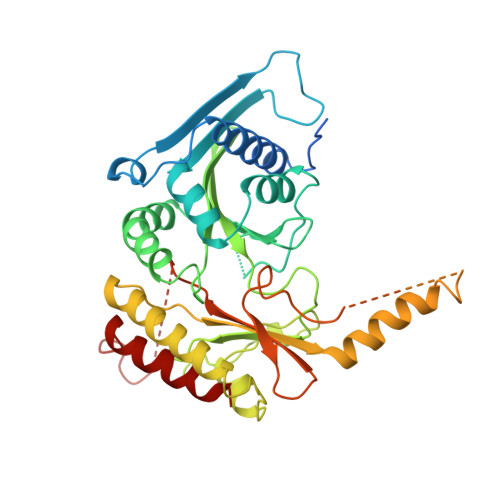
Structure of type IIbeta phosphatidylinositol phosphate kinase: a protein kinase fold flattened for interfacial phosphorylation.

Rao, V.D.Misra, S.Boronenkov, I.V.Anderson, R.A.Hurley, J.H.

(1998) Cell 94: 829-839

  • DOI: https://doi.org/10.1016/s0092-8674(00)81741-9
  • Primary Citation Related Structures: 
    1BO1

  • PubMed Abstract: 

    Phosphoinositide kinases play central roles in signal transduction by phosphorylating the inositol ring at specific positions. The structure of one such enzyme, type IIbeta phosphatidylinositol phosphate kinase, reveals a protein kinase ATP-binding core and demonstrates that all phosphoinositide kinases belong to one superfamily. The enzyme is a disc-shaped homodimer with a 33 x 48 A basic flat face that suggests an electrostatic mechanism for plasma membrane targeting. Conserved basic residues form a putative phosphatidylinositol phosphate specificity site. The substrate-binding site is open on one side, consistent with dual specificity for phosphatidylinositol 3- and 5-phosphates. A modeled complex with membrane-bound substrate and ATP shows how a phosphoinositide kinase can phosphorylate its substrate in situ at the membrane interface.


  • Organizational Affiliation
    • Laboratory of Molecular Biology, National Institute of Diabetes, Digestive and Kidney Diseases, National Institutes of Health, Bethesda, Maryland 20892-0580, USA.

Macromolecule Content 

  • Total Structure Weight: 94.9 kDa 
  • Atom Count: 5,301 
  • Modeled Residue Count: 644 
  • Deposited Residue Count: 832 
  • Unique protein chains: 1

Macromolecules

Find similar proteins by:|  3D Structure
Entity ID: 1
MoleculeChains  Sequence LengthOrganismDetailsImage
PROTEIN (PHOSPHATIDYLINOSITOL PHOSPHATE KINASE IIBETA)
A, B
416Homo sapiensMutation(s): 0 
EC: 2.7.1.68 (PDB Primary Data), 2.7.1.149 (UniProt)
UniProt & NIH Common Fund Data Resources
Find proteins for P78356 (Homo sapiens)
Explore P78356 
Go to UniProtKB:  P78356
PHAROS:  P78356
GTEx:  ENSG00000276293 
Entity Groups
Sequence Clusters30% Identity50% Identity70% Identity90% Identity95% Identity100% Identity
UniProt GroupP78356
Sequence Annotations
Expand
Reference Sequence

Experimental Data & Validation

Experimental Data

  • Method: X-RAY DIFFRACTION
  • Resolution: 3.00 Å
  • R-Value Free:  0.299 (Depositor) 
  • R-Value Work:  0.229 (Depositor) 
  • R-Value Observed: 0.229 (Depositor) 
Space Group: C 2 2 21
Unit Cell:
Length ( Å )Angle ( ˚ )
a = 109.915α = 90
b = 182.403β = 90
c = 106.472γ = 90
Software Package:
Software NamePurpose
DENZOdata reduction
SCALEPACKdata scaling
SHARPphasing
X-PLORrefinement

Structure Validation

View Full Validation Report


Entry History 

Deposition Data

Revision History  (Full details and data files)

  • Version 1.0: 1998-10-07
    Type: Initial release
  • Version 1.1: 2007-10-16
    Changes: Version format compliance
  • Version 1.2: 2011-07-13
    Changes: Version format compliance
  • Version 1.3: 2024-02-07
    Changes: Data collection, Database references