1BGJ

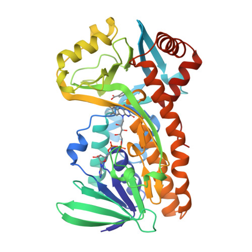
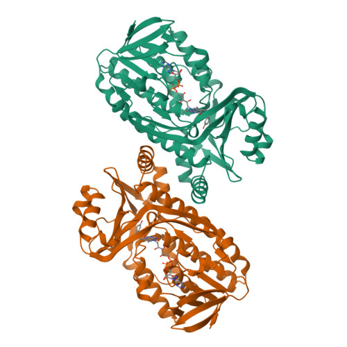
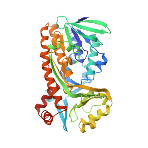
P-HYDROXYBENZOATE HYDROXYLASE (PHBH) MUTANT WITH CYS 116 REPLACED BY SER (C116S) AND HIS 162 REPLACED BY ARG (H162R), IN COMPLEX WITH FAD AND 4-HYDROXYBENZOIC ACID


Experimental Data Snapshot

  • Method: X-RAY DIFFRACTION
  • Resolution: 3.00 Å
  • R-Value Work: 
    0.128 (Depositor), 0.130 (DCC) 
  • R-Value Observed: 
    0.128 (Depositor) 

Starting Model: experimental
View more details

wwPDB Validation   3D Report Full Report


Ligand Structure Quality Assessment 

Created with Raphaël 2.3.0Worse 01 BetterLigand structure goodness of fit to experimental dataBest fitted FADClick on this verticalbar to view details

This is version 1.7 of the entry. See complete history


Literature

Interdomain binding of NADPH in p-hydroxybenzoate hydroxylase as suggested by kinetic, crystallographic and modeling studies of histidine 162 and arginine 269 variants.

Eppink, M.H.Schreuder, H.A.van Berkel, W.J.

(1998) J Biol Chem 273: 21031-21039

  • DOI: https://doi.org/10.1074/jbc.273.33.21031
  • Primary Citation of Related Structures:  
    1BGJ, 1BGN

  • PubMed Abstract: 

    The conserved residues His-162 and Arg-269 of the flavoprotein p-hydroxybenzoate hydroxylase (EC 1.14.13.2) are located at the entrance of the interdomain cleft that leads toward the active site. To study their putative role in NADPH binding, His-162 and Arg-269 were selectively changed by site-specific mutagenesis. The catalytic properties of H162R, H162Y, and R269K were similar to the wild-type enzyme. However, less conservative His-162 and Arg-269 replacements strongly impaired NADPH binding without affecting the conformation of the flavin ring and the efficiency of substrate hydroxylation. The crystal structures of H162R and R269T in complex with 4-hydroxybenzoate were solved at 3.0 and 2.0 A resolution, respectively. Both structures are virtually indistinguishable from the wild-type enzyme-substrate complex except for the substituted side chains. In contrast to wild-type p-hydroxybenzoate hydroxylase, H162R is not inactivated by diethyl pyrocarbonate. NADPH protects wild-type p-hydroxybenzoate hydroxylase from diethylpyrocarbonate inactivation, suggesting that His-162 is involved in NADPH binding. Based on these results and GRID calculations we propose that the side chains of His-162 and Arg-269 interact with the pyrophosphate moiety of NADPH. An interdomain binding mode for NADPH is proposed which takes a novel sequence motif (Eppink, M. H. M., Schreuder, H. A., and van Berkel, W. J. H. (1997) Protein Sci. 6, 2454-2458) into account.


  • Organizational Affiliation

    Department of Biomolecular Sciences, Laboratory of Biochemistry, Wageningen Agricultural University, Dreijenlaan 3, 6703 HA Wageningen, The Netherlands.


Macromolecules
Find similar proteins by:  (by identity cutoff)  |  3D Structure
Entity ID: 1
MoleculeChains Sequence LengthOrganismDetailsImage
P-HYDROXYBENZOATE HYDROXYLASE394Pseudomonas fluorescensMutation(s): 2 
Gene Names: POBA
EC: 1.14.13.2
UniProt
Find proteins for P00438 (Pseudomonas fluorescens)
Explore P00438 
Go to UniProtKB:  P00438
Entity Groups  
Sequence Clusters30% Identity50% Identity70% Identity90% Identity95% Identity100% Identity
UniProt GroupP00438
Sequence Annotations
Expand
  • Reference Sequence
Experimental Data & Validation

Experimental Data

  • Method: X-RAY DIFFRACTION
  • Resolution: 3.00 Å
  • R-Value Work:  0.128 (Depositor), 0.130 (DCC) 
  • R-Value Observed: 0.128 (Depositor) 
Space Group: C 2 2 21
Unit Cell:
Length ( Å )Angle ( ˚ )
a = 71.8α = 90
b = 146β = 90
c = 88.3γ = 90
Software Package:
Software NamePurpose
X-PLORmodel building
X-PLORrefinement
XDSdata reduction
XSCALEdata scaling
X-PLORphasing

Structure Validation

View Full Validation Report


Ligand Structure Quality Assessment 

Created with Raphaël 2.3.0Worse 01 BetterLigand structure goodness of fit to experimental dataBest fitted FADClick on this verticalbar to view details

Entry History 

Deposition Data

Revision History  (Full details and data files)

  • Version 1.0: 1998-08-12
    Type: Initial release
  • Version 1.1: 2008-03-24
    Changes: Version format compliance
  • Version 1.2: 2011-07-13
    Changes: Derived calculations, Version format compliance
  • Version 1.3: 2017-11-29
    Changes: Derived calculations, Other
  • Version 1.4: 2018-04-04
    Changes: Data collection
  • Version 1.5: 2021-11-03
    Changes: Database references, Derived calculations
  • Version 1.6: 2023-08-02
    Changes: Refinement description
  • Version 1.7: 2024-05-22
    Changes: Data collection