1BF4 | pdb_00001bf4

CHROMOSOMAL DNA-BINDING PROTEIN SSO7D/D(GCGAACGC) COMPLEX


Experimental Data Snapshot

  • Method: X-RAY DIFFRACTION
  • Resolution: 1.60 Å
  • R-Value Free: 
    0.288 (Depositor) 
  • R-Value Work: 
    0.211 (Depositor), 0.214 (DCC) 
  • R-Value Observed: 
    0.211 (Depositor) 

Starting Model: experimental
View more details

wwPDB Validation 3D Report Full Report

Validation slider image for 1BF4

This is version 1.5 of the entry. See complete history

Literature

The crystal structure of the hyperthermophile chromosomal protein Sso7d bound to DNA.

Gao, Y.G.Su, S.Y.Robinson, H.Padmanabhan, S.Lim, L.McCrary, B.S.Edmondson, S.P.Shriver, J.W.Wang, A.H.

(1998) Nat Struct Biol 5: 782-786

  • DOI: https://doi.org/10.1038/1822
  • Primary Citation Related Structures: 
    1BF4, 1BNZ

  • PubMed Abstract: 

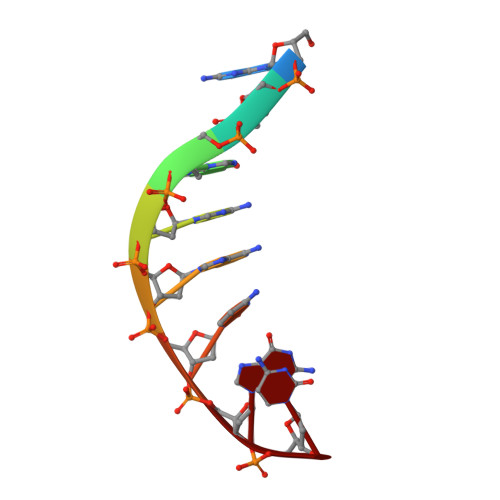
    Sso7d and Sac7d are two small (approximately 7,000 Mr), but abundant, chromosomal proteins from the hyperthermophilic archaeabacteria Sulfolobus solfataricus and S. acidocaldarius respectively. These proteins have high thermal, acid and chemical stability. They bind DNA without marked sequence preference and increase the Tm of DNA by approximately 40 degrees C. Sso7d in complex with GTAATTAC and GCGT(iU)CGC + GCGAACGC was crystallized in different crystal lattices and the crystal structures were solved at high resolution. Sso7d binds in the minor groove of DNA and causes a single-step sharp kink in DNA (approximately 60 degrees) by the intercalation of the hydrophobic side chains of Val 26 and Met 29. The intercalation sites are different in the two complexes. Observations of this novel DNA binding mode in three independent crystal lattices indicate that it is not a function of crystal packing.


  • Organizational Affiliation
    • Department of Cell and Structural Biology, University of Illinois at Urbana-Champaign, Urbana 61801, USA.

Macromolecule Content 

  • Total Structure Weight: 12.13 kDa 
  • Atom Count: 938 
  • Modeled Residue Count: 79 
  • Deposited Residue Count: 79 
  • Unique protein chains: 1
  • Unique nucleic acid chains: 2

Macromolecules


Find similar proteins by:|  3D Structure
Entity ID: 3
MoleculeChains  Sequence LengthOrganismDetailsImage
PROTEIN (CHROMOSOMAL PROTEIN SSO7D)C [auth A]63Sulfolobus acidocaldariusMutation(s): 0 
UniProt
Find proteins for P13123 (Sulfolobus acidocaldarius (strain ATCC 33909 / DSM 639 / JCM 8929 / NBRC 15157 / NCIMB 11770))
Explore P13123 
Go to UniProtKB:  P13123
Entity Groups
Sequence Clusters30% Identity50% Identity70% Identity90% Identity95% Identity100% Identity
UniProt GroupP13123
Sequence Annotations
Expand
Reference Sequence
Find similar nucleic acids by:  Sequence
Entity ID: 1
MoleculeChains LengthOrganismImage
DNA (5'-D(*GP*CP*GP*TP*5IUP*CP*GP*C)-3')A [auth B]8N/A
Sequence Annotations
Expand
Reference Sequence
Find similar nucleic acids by:  Sequence
Entity ID: 2
MoleculeChains LengthOrganismImage
DNA (5'-D(*GP*CP*GP*AP*AP*CP*GP*C)-3')B [auth C]8N/A
Sequence Annotations
Expand
Reference Sequence

Experimental Data & Validation

Experimental Data

  • Method: X-RAY DIFFRACTION
  • Resolution: 1.60 Å
  • R-Value Free:  0.288 (Depositor) 
  • R-Value Work:  0.211 (Depositor), 0.214 (DCC) 
  • R-Value Observed: 0.211 (Depositor) 
Space Group: P 21 21 21
Unit Cell:
Length ( Å )Angle ( ˚ )
a = 46.87α = 90
b = 49.67β = 90
c = 37.65γ = 90
Software Package:
Software NamePurpose
AMoREphasing
X-PLORrefinement
bioteXdata reduction
bioteXdata scaling

Structure Validation

View Full Validation Report


Entry History 

Deposition Data

Revision History  (Full details and data files)

  • Version 1.0: 1999-11-10
    Type: Initial release
  • Version 1.1: 2008-05-22
    Changes: Version format compliance
  • Version 1.2: 2011-07-13
    Changes: Version format compliance
  • Version 1.3: 2017-11-22
    Changes: Refinement description
  • Version 1.4: 2024-02-07
    Changes: Data collection, Database references, Derived calculations
  • Version 1.5: 2024-04-03
    Changes: Refinement description