1BDW | pdb_00001bdw

GRAMICIDIN D FROM BACILLUS BREVIS (ACTIVE FORM)


Experimental Data Snapshot

  • Method: X-RAY DIFFRACTION
  • Resolution: 1.70 Å
  • R-Value Free: 
    0.243 (Depositor), 0.195 (DCC) 
  • R-Value Work: 
    0.187 (DCC) 
  • R-Value Observed: 
    0.168 (Depositor) 

Starting Model: experimental
View more details

wwPDB Validation 3D Report Full Report

Validation slider image for 1BDW

This is version 2.1 of the entry. See complete history

Literature

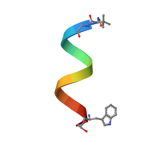
The Conducting Form of Gramicidin a is a Right-Handed Double-Stranded Double Helix.

Burkhart, B.M.Li, N.Langs, D.A.Pangborn, W.A.Duax, W.L.

(1998) Proc Natl Acad Sci U S A 95: 12950

  • DOI: https://doi.org/10.1073/pnas.95.22.12950
  • Primary Citation Related Structures: 
    1AV2, 1BDW

  • PubMed Abstract: 

    The linear pentadecapeptide antibiotic, gramicidin D, is a naturally occurring product of Bacillus brevis known to form ion channels in synthetic and natural membranes. The x-ray crystal structures of the right-handed double-stranded double-helical dimers (DSDH) reported here agree with 15N-NMR and CD data on the functional gramicidin D channel in lipid bilayers. These structures demonstrate single-file ion transfer through the channels. The results also indicate that previous crystal structure reports of a left-handed double-stranded double-helical dimer in complex with Cs+ and K+ salts may be in error and that our evidence points to the DSDH as the major conformer responsible for ion transport in membranes.


  • Organizational Affiliation
    • Hauptman-Woodward Medical Research Institute, Inc., 73 High Street, Buffalo, NY 14203, USA. burkhart@hwi.buffalo.edu

Macromolecule Content 

  • Total Structure Weight: 4.06 kDa 
  • Atom Count: 338 
  • Modeled Residue Count: 32 
  • Deposited Residue Count: 32 
  • Unique protein chains: 1

Macromolecules

Find similar proteins by:  Sequence   |   3D Structure  
Entity ID: 1
MoleculeChains  Sequence LengthOrganismDetailsImage
GRAMICIDIN A
A, B
16Brevibacillus brevisMutation(s): 0 

Biologically Interesting Molecules (External Reference) 

1 Unique

Experimental Data & Validation

Experimental Data

  • Method: X-RAY DIFFRACTION
  • Resolution: 1.70 Å
  • R-Value Free:  0.243 (Depositor), 0.195 (DCC) 
  • R-Value Work:  0.187 (DCC) 
  • R-Value Observed: 0.168 (Depositor) 
Space Group: P 21 21 21
Unit Cell:
Length ( Å )Angle ( ˚ )
a = 20.584α = 90
b = 27.905β = 90
c = 52.04γ = 90
Software Package:
Software NamePurpose
AMoREphasing
SHELXL-97refinement
DENZOdata reduction
SCALEPACKdata scaling

Structure Validation

View Full Validation Report


Entry History 

Deposition Data

Revision History  (Full details and data files)

  • Version 1.0: 1998-10-14
    Type: Initial release
  • Version 1.1: 2011-06-14
    Changes: Version format compliance
  • Version 1.2: 2011-07-13
    Changes: Version format compliance
  • Version 1.3: 2011-07-27
    Changes: Atomic model, Database references, Derived calculations, Non-polymer description, Structure summary
  • Version 1.4: 2012-12-12
    Changes: Other
  • Version 1.5: 2023-08-02
    Changes: Database references, Derived calculations, Refinement description
  • Version 2.0: 2023-11-15
    Changes: Atomic model, Data collection, Derived calculations
  • Version 2.1: 2024-10-30
    Changes: Structure summary