1BBS | pdb_00001bbs

X-RAY ANALYSES OF PEPTIDE INHIBITOR COMPLEXES DEFINE THE STRUCTURAL BASIS OF SPECIFICITY FOR HUMAN AND MOUSE RENINS


Experimental Data Snapshot

  • Method: X-RAY DIFFRACTION
  • Resolution: 2.80 Å
  • R-Value Observed: 
    0.196 (Depositor) 

wwPDB Validation 3D Report Full Report

Validation slider image for 1BBS

This is version 1.4 of the entry. See complete history

Literature

X-ray analyses of peptide-inhibitor complexes define the structural basis of specificity for human and mouse renins.

Dhanaraj, V.Dealwis, C.G.Frazao, C.Badasso, M.Sibanda, B.L.Tickle, I.J.Cooper, J.B.Driessen, H.P.Newman, M.Aguilar, C.Wood, S.P.Blundell, T.L.Hobart, P.M.Geoghegan, K.F.Ammirati, M.J.Danley, D.E.

(1992) Nature 357: 466-472

  • DOI: https://doi.org/10.1038/357466a0
  • Primary Citation Related Structures: 
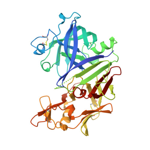
    1BBS

  • PubMed Abstract: 

    X-ray analyses have defined the three-dimensional structures of crystals of mouse and human renins complexed with peptide inhibitors at resolutions of 1.9 and 2.8 A, respectively. The exquisite specificity of renin arises partly from ordered loop regions at the periphery of the binding cleft. Although the pattern of main-chain hydrogen bonding in other aspartic proteinase inhibitor complexes is conserved in renins, differences in the positions of secondary structure elements (particularly helices) also lead to improved specificity in renins for angiotensinogen substrates.


  • Organizational Affiliation
    • Department of Crystallography, Birkbeck College, London, UK.

Macromolecule Content 

  • Total Structure Weight: 74.53 kDa 
  • Atom Count: 5,018 
  • Modeled Residue Count: 662 
  • Deposited Residue Count: 680 
  • Unique protein chains: 1

Macromolecules

Find similar proteins by:|  3D Structure
Entity ID: 1
MoleculeChains  Sequence LengthOrganismDetailsImage
RENIN
A, B
340Homo sapiensMutation(s): 0 
EC: 3.4.23.15
UniProt & NIH Common Fund Data Resources
Find proteins for P00797 (Homo sapiens)
Explore P00797 
Go to UniProtKB:  P00797
PHAROS:  P00797
GTEx:  ENSG00000143839 
Entity Groups
Sequence Clusters30% Identity50% Identity70% Identity90% Identity95% Identity100% Identity
UniProt GroupP00797
Sequence Annotations
Expand
Reference Sequence

Experimental Data & Validation

Experimental Data

  • Method: X-RAY DIFFRACTION
  • Resolution: 2.80 Å
  • R-Value Observed: 0.196 (Depositor) 
Space Group: P 21 3
Unit Cell:
Length ( Å )Angle ( ˚ )
a = 143.1α = 90
b = 143.1β = 90
c = 143.1γ = 90
Software Package:
Software NamePurpose
RESTRAINrefinement

Structure Validation

View Full Validation Report


Entry History 

Deposition Data

Revision History  (Full details and data files)

  • Version 1.0: 1994-01-31
    Type: Initial release
  • Version 1.1: 2008-03-03
    Changes: Version format compliance
  • Version 1.2: 2011-07-13
    Changes: Version format compliance
  • Version 1.3: 2017-11-29
    Changes: Derived calculations, Other
  • Version 1.4: 2024-11-06
    Changes: Data collection, Database references, Structure summary