1BA7 | pdb_00001ba7

SOYBEAN TRYPSIN INHIBITOR


Experimental Data Snapshot

  • Method: X-RAY DIFFRACTION
  • Resolution: 2.50 Å
  • R-Value Free: 
    0.223 (Depositor) 
  • R-Value Work: 
    0.160 (Depositor), 0.168 (DCC) 
  • R-Value Observed: 
    0.160 (Depositor) 

Starting Model: experimental
View more details

wwPDB Validation 3D Report Full Report

Validation slider image for 1BA7

This is version 1.5 of the entry. See complete history

Literature

Structure of the Kunitz-type soybean trypsin inhibitor (STI): implication for the interactions between members of the STI family and tissue-plasminogen activator.

De Meester, P.Brick, P.Lloyd, L.F.Blow, D.M.Onesti, S.

(1998) Acta Crystallogr D Biol Crystallogr 54: 589-597

  • DOI: https://doi.org/10.1107/s0907444997015849
  • Primary Citation Related Structures: 
    1BA7

  • PubMed Abstract: 

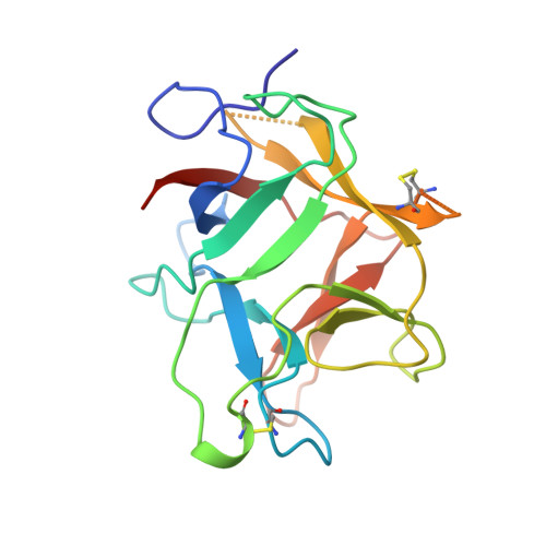
    The Kunitz-type soybean trypsin inhibitor (STI) has played a key role in the early study of proteinases, having been used as the main substrate in the biochemical and kinetic work that led to the definition of the standard mechanism of action of proteinase inhibitors. A partial structure of STI complexed with porcine trypsin has previously been reported, in which the first 93 residues of the inhibitor, including the region of contact with trypsin, were relatively well defined, whereas for the remaining part of the peptide chain only some Calpha atoms were located. The structure of the inhibitor in its free form has now been determined by molecular replacement to 2.5 A, using the coordinates of the homologous Erythrina trypsin inhibitor as a search model. When the refined atomic coordinates of STI are compared with the partial model previously available, the conformation of the reactive-site loop and its position with respect to the main body of the molecule does not change when the inhibitor interacts with trypsin. There are instead, despite the high similarity in the overall tertiary structure, significant differences between STI and Erythrina trypsin inhibitor (ETI) in the region which is in contact with the enzyme in the STI:trypsin crystal structure. Some of these differences can explain the unique specificity of ETI and its ability to inhibit the fibrinolytic enzyme tissue-type plasminogen activator.


  • Organizational Affiliation
    • Blackett Laboratory, Imperial College, London SW7 2BZ, England.

Macromolecule Content 

  • Total Structure Weight: 40.23 kDa 
  • Atom Count: 2,653 
  • Modeled Residue Count: 334 
  • Deposited Residue Count: 362 
  • Unique protein chains: 1

Macromolecules

Find similar proteins by:|  3D Structure
Entity ID: 1
MoleculeChains  Sequence LengthOrganismDetailsImage
TRYPSIN INHIBITOR (KUNITZ)
A, B
181Glycine maxMutation(s): 0 
UniProt
Find proteins for P01070 (Glycine max)
Explore P01070 
Go to UniProtKB:  P01070
Entity Groups
Sequence Clusters30% Identity50% Identity70% Identity90% Identity95% Identity100% Identity
UniProt GroupP01070
Sequence Annotations
Expand
Reference Sequence

Experimental Data & Validation

Experimental Data

  • Method: X-RAY DIFFRACTION
  • Resolution: 2.50 Å
  • R-Value Free:  0.223 (Depositor) 
  • R-Value Work:  0.160 (Depositor), 0.168 (DCC) 
  • R-Value Observed: 0.160 (Depositor) 
Space Group: P 1
Unit Cell:
Length ( Å )Angle ( ˚ )
a = 39.11α = 71.75
b = 53.87β = 90
c = 58.2γ = 82.98
Software Package:
Software NamePurpose
MADNESdata collection
CCP4data reduction
X-PLORmodel building
X-PLORrefinement
MADNESdata reduction
CCP4data scaling
X-PLORphasing

Structure Validation

View Full Validation Report


Entry History 

Deposition Data

Revision History  (Full details and data files)

  • Version 1.0: 1998-06-17
    Type: Initial release
  • Version 1.1: 2008-03-24
    Changes: Version format compliance
  • Version 1.2: 2011-07-13
    Changes: Version format compliance
  • Version 1.3: 2018-04-18
    Changes: Data collection, Other
  • Version 1.4: 2023-08-02
    Changes: Database references, Refinement description
  • Version 1.5: 2024-11-06
    Changes: Data collection, Structure summary