1B6T | pdb_00001b6t

PHOSPHOPANTETHEINE ADENYLYLTRANSFERASE IN COMPLEX WITH 3'-DEPHOSPHO-COA FROM ESCHERICHIA COLI


Experimental Data Snapshot

  • Method: X-RAY DIFFRACTION
  • Resolution: 1.80 Å
  • R-Value Free: 
    0.235 (Depositor) 
  • R-Value Work: 
    0.205 (Depositor) 
  • R-Value Observed: 
    0.222 (Depositor) 

wwPDB Validation 3D Report Full Report

Validation slider image for 1B6T

This is version 1.3 of the entry. See complete history

Literature

The crystal structure of a novel bacterial adenylyltransferase reveals half of sites reactivity.

Izard, T.Geerlof, A.

(1999) EMBO J 18: 2021-2030

  • DOI: https://doi.org/10.1093/emboj/18.8.2021
  • Primary Citation Related Structures: 
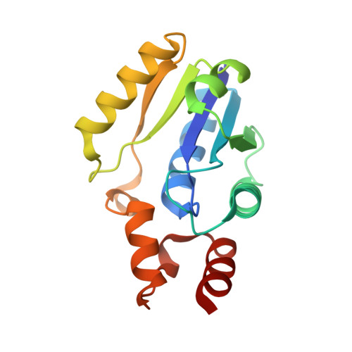
    1B6T

  • PubMed Abstract: 

    Phosphopantetheine adenylyltransferase (PPAT) is an essential enzyme in bacteria that catalyses a rate-limiting step in coenzyme A (CoA) biosynthesis, by transferring an adenylyl group from ATP to 4'-phosphopantetheine, yielding dephospho-CoA (dPCoA). Each phosphopantetheine adenylyltransferase (PPAT) subunit displays a dinucleotide-binding fold that is structurally similar to that in class I aminoacyl-tRNA synthetases. Superposition of bound adenylyl moieties from dPCoA in PPAT and ATP in aminoacyl-tRNA synthetases suggests nucleophilic attack by the 4'-phosphopantetheine on the alpha-phosphate of ATP. The proposed catalytic mechanism implicates transition state stabilization by PPAT without involving functional groups of the enzyme in a chemical sense in the reaction. The crystal structure of the enzyme from Escherichia coli in complex with dPCoA shows that binding at one site causes a vice-like movement of active site residues lining the active site surface. The mode of enzyme product formation is highly concerted, with only one trimer of the PPAT hexamer showing evidence of dPCoA binding. The homologous active site attachment of ATP and the structural distribution of predicted sequence-binding motifs in PPAT classify the enzyme as belonging to the nucleotidyltransferase superfamily.


  • Organizational Affiliation
    • Department of Biochemistry, University of Leicester, Leicester LE1 7RH.ti3@le.ac.uk

Macromolecule Content 

  • Total Structure Weight: 36.69 kDa 
  • Atom Count: 2,853 
  • Modeled Residue Count: 314 
  • Deposited Residue Count: 318 
  • Unique protein chains: 1

Macromolecules

Find similar proteins by:|  3D Structure
Entity ID: 1
MoleculeChains  Sequence LengthOrganismDetailsImage
PROTEIN (PHOSPHOPANTETHEINE ADENYLYLTRANSFERASE)
A, B
159Escherichia coliMutation(s): 0 
EC: 2.7.7.3
UniProt
Find proteins for P0A6I6 (Escherichia coli (strain K12))
Explore P0A6I6 
Go to UniProtKB:  P0A6I6
Entity Groups
Sequence Clusters30% Identity50% Identity70% Identity90% Identity95% Identity100% Identity
UniProt GroupP0A6I6
Sequence Annotations
Expand
Reference Sequence

Experimental Data & Validation

Experimental Data

  • Method: X-RAY DIFFRACTION
  • Resolution: 1.80 Å
  • R-Value Free:  0.235 (Depositor) 
  • R-Value Work:  0.205 (Depositor) 
  • R-Value Observed: 0.222 (Depositor) 
Space Group: I 2 3
Unit Cell:
Length ( Å )Angle ( ˚ )
a = 135.5α = 90
b = 135.5β = 90
c = 135.5γ = 90
Software Package:
Software NamePurpose
CNSrefinement
DENZOdata reduction
SCALEPACKdata scaling

Structure Validation

View Full Validation Report


Entry History 

Deposition Data

  • Released Date: 2000-04-19 
  • Deposition Author(s): Izard, T.

Revision History  (Full details and data files)

  • Version 1.0: 2000-04-19
    Type: Initial release
  • Version 1.1: 2008-04-27
    Changes: Version format compliance
  • Version 1.2: 2011-07-13
    Changes: Version format compliance
  • Version 1.3: 2023-12-27
    Changes: Data collection, Database references, Derived calculations