1B6Q | pdb_00001b6q

ALANINE 31 PROLINE MUTANT OF ROP PROTEIN


Experimental Data Snapshot

  • Method: X-RAY DIFFRACTION
  • Resolution: 1.80 Å
  • R-Value Free: 
    0.240 (Depositor) 
  • R-Value Work: 
    0.189 (Depositor) 
  • R-Value Observed: 
    0.189 (Depositor) 

Starting Model: experimental
View more details

wwPDB Validation 3D Report Full Report

Validation slider image for 1B6Q

This is version 1.7 of the entry. See complete history

Literature

Protein plasticity to the extreme: changing the topology of a 4-alpha-helical bundle with a single amino acid substitution.

Glykos, N.M.Cesareni, G.Kokkinidis, M.

(1999) Structure 7: 597-603

  • DOI: https://doi.org/10.1016/s0969-2126(99)80081-1
  • Primary Citation Related Structures: 
    1B6Q

  • PubMed Abstract: 

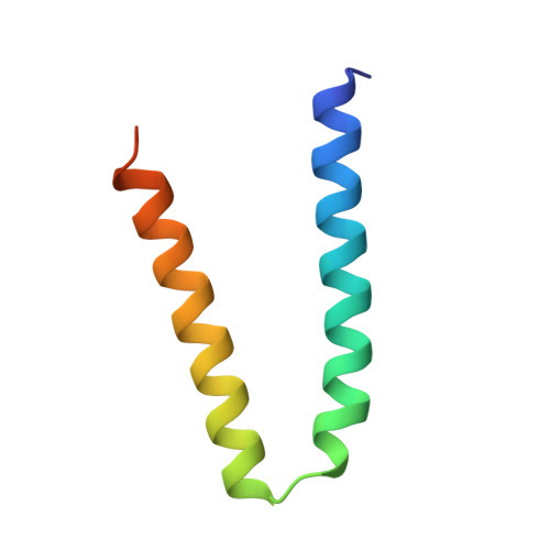
    Conventional wisdom has it that two proteins sharing 98.4% sequence identity have nearly identical three-dimensional structures. Here we provide a counter-example to this statement by showing that a single amino acid substitution can change the topology of a homodimeric 4-alpha-helical bundle protein. We have determined the high-resolution crystal structure of a 4-alpha-helical protein with a single alanine to proline mutation in the turn region, and show that this single amino acid substitution leads to a complete reorganisation of the whole molecule. The protein is converted from the canonical left-handed all-antiparallel form, to a right-handed mixed parallel and antiparallel bundle, which to the best of our knowledge and belief represents a novel topological motif for this class of proteins. The results suggest a possible new mechanism for the creation and evolution of topological motifs, show the importance of loop regions in determining the allowable folding pathways, and illustrate the malleability of protein structures.


  • Organizational Affiliation
    • Foundation for Research and Technology-Hellas, Institute of Molecular Biology and Biotechnology, Heraklion, Crete, Greece.

Macromolecule Content 

  • Total Structure Weight: 7.26 kDa 
  • Atom Count: 544 
  • Modeled Residue Count: 56 
  • Deposited Residue Count: 63 
  • Unique protein chains: 1

Macromolecules

Find similar proteins by:|  3D Structure
Entity ID: 1
MoleculeChains  Sequence LengthOrganismDetailsImage
ROP63Escherichia coliMutation(s): 1 
Gene Names: ROP
UniProt
Find proteins for P03051 (Escherichia coli)
Explore P03051 
Go to UniProtKB:  P03051
Entity Groups
Sequence Clusters30% Identity50% Identity70% Identity90% Identity95% Identity100% Identity
UniProt GroupP03051
Sequence Annotations
Expand
Reference Sequence

Experimental Data & Validation

Experimental Data

  • Method: X-RAY DIFFRACTION
  • Resolution: 1.80 Å
  • R-Value Free:  0.240 (Depositor) 
  • R-Value Work:  0.189 (Depositor) 
  • R-Value Observed: 0.189 (Depositor) 
Space Group: C 2 2 21
Unit Cell:
Length ( Å )Angle ( ˚ )
a = 30.4α = 90
b = 42.1β = 90
c = 81.4γ = 90
Software Package:
Software NamePurpose
X-PLORmodel building
X-PLORrefinement
CAD4data reduction
CAD4data scaling
X-PLORphasing

Structure Validation

View Full Validation Report


Entry History 

Deposition Data

Revision History  (Full details and data files)

  • Version 1.0: 1999-07-09
    Type: Initial release
  • Version 1.1: 2008-03-24
    Changes: Version format compliance
  • Version 1.2: 2011-07-13
    Changes: Derived calculations, Version format compliance
  • Version 1.3: 2011-11-16
    Changes: Atomic model
  • Version 1.4: 2018-04-04
    Changes: Data collection, Other
  • Version 1.5: 2018-04-18
    Changes: Data collection
  • Version 1.6: 2023-08-02
    Changes: Database references, Refinement description
  • Version 1.7: 2024-05-22
    Changes: Data collection