1B04 | pdb_00001b04

STRUCTURE OF THE ADENYLATION DOMAIN OF AN NAD+ DEPENDENT LIGASE


Experimental Data Snapshot

  • Method: X-RAY DIFFRACTION
  • Resolution: 2.80 Å
  • R-Value Free: 
    0.310 (Depositor), 0.299 (DCC) 
  • R-Value Work: 
    0.230 (Depositor), 0.226 (DCC) 
  • R-Value Observed: 
    0.230 (Depositor) 

wwPDB Validation 3D Report Full Report

Validation slider image for 1B04

This is version 1.4 of the entry. See complete history

Literature

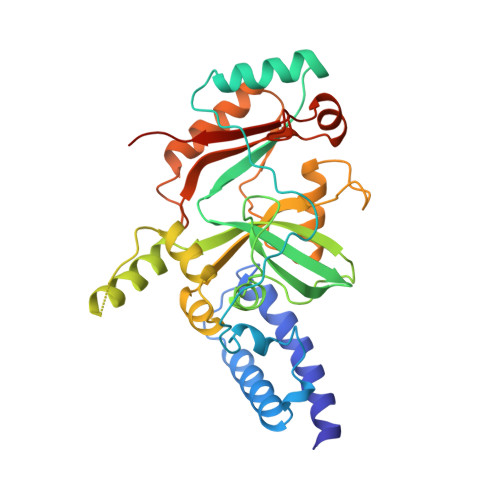
Structure of the adenylation domain of an NAD+-dependent DNA ligase.

Singleton, M.R.Hakansson, K.Timson, D.J.Wigley, D.B.

(1999) Structure 7: 35-42

  • DOI: https://doi.org/10.1016/s0969-2126(99)80007-0
  • Primary Citation Related Structures: 
    1B04

  • PubMed Abstract: 

    DNA ligases catalyse phosphodiester bond formation between adjacent bases in nicked DNA, thereby sealing the nick. A key step in the catalytic mechanism is the formation of an adenylated DNA intermediate. The adenyl group is derived from either ATP (in eucaryotes and archaea) or NAD+4 (in bacteria). This difference in cofactor specificity suggests that DNA ligase may be a useful antibiotic target. The crystal structure of the adenylation domain of the NAD+-dependent DNA ligase from Bacillus stearothermophilus has been determined at 2.8 A resolution. Despite a complete lack of detectable sequence similarity, the fold of the central core of this domain shares homology with the equivalent region of ATP-dependent DNA ligases, providing strong evidence for the location of the NAD+-binding site. Comparison of the structure of the NAD+4-dependent DNA ligase with that of ATP-dependent ligases and mRNA-capping enzymes demonstrates the manifold utilisation of a conserved nucleotidyltransferase domain within this family of enzymes. Whilst this conserved core domain retains a common mode of nucleotide binding and activation, it is the additional domains at the N terminus and/or the C terminus that provide the alternative specificities and functionalities in the different members of this enzyme superfamily.


  • Organizational Affiliation
    • Sir William Dunn School of Pathology, University of Oxford, South ParksRoad, Oxford OX1 3RE, UK.

Macromolecule Content 

  • Total Structure Weight: 71.66 kDa 
  • Atom Count: 5,126 
  • Modeled Residue Count: 621 
  • Deposited Residue Count: 636 
  • Unique protein chains: 1

Macromolecules

Find similar proteins by:|  3D Structure
Entity ID: 1
MoleculeChains  Sequence LengthOrganismDetailsImage
PROTEIN (DNA LIGASE)
A, B
318Geobacillus stearothermophilusMutation(s): 1 
Gene Names: LIG
EC: 6.5.1.2
UniProt
Find proteins for O87703 (Geobacillus stearothermophilus)
Explore O87703 
Go to UniProtKB:  O87703
Entity Groups
Sequence Clusters30% Identity50% Identity70% Identity90% Identity95% Identity100% Identity
UniProt GroupO87703
Sequence Annotations
Expand
Reference Sequence

Experimental Data & Validation

Experimental Data

  • Method: X-RAY DIFFRACTION
  • Resolution: 2.80 Å
  • R-Value Free:  0.310 (Depositor), 0.299 (DCC) 
  • R-Value Work:  0.230 (Depositor), 0.226 (DCC) 
  • R-Value Observed: 0.230 (Depositor) 
Space Group: P 43 21 2
Unit Cell:
Length ( Å )Angle ( ˚ )
a = 95.72α = 90
b = 95.72β = 90
c = 225.89γ = 90
Software Package:
Software NamePurpose
REFMACrefinement
DENZOdata reduction
SCALEPACKdata scaling

Structure Validation

View Full Validation Report


Entry History 

Deposition Data

Revision History  (Full details and data files)

  • Version 1.0: 1999-11-22
    Type: Initial release
  • Version 1.1: 2008-04-27
    Changes: Version format compliance
  • Version 1.2: 2011-07-13
    Changes: Version format compliance
  • Version 1.3: 2021-11-03
    Changes: Data collection, Database references
  • Version 1.4: 2024-02-07
    Changes: Data collection