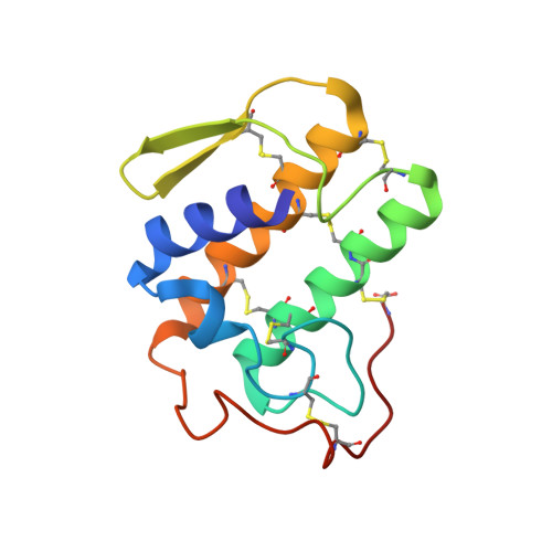
1AYP

A PROBE MOLECULE COMPOSED OF SEVENTEEN PERCENT OF TOTAL DIFFRACTING MATTER GIVES CORRECT SOLUTIONS IN MOLECULAR REPLACEMENT


Experimental Data Snapshot

  • Method: X-RAY DIFFRACTION
  • Resolution: 2.57 Å
  • R-Value Work: 0.214 
  • R-Value Observed: 0.214 

wwPDB Validation   3D Report Full Report


Ligand Structure Quality Assessment 


This is version 1.4 of the entry. See complete history


Literature

A probe molecule composed of seventeen percent of total diffracting matter gives correct solutions in molecular replacement.

Oh, B.H.

(1995) Acta Crystallogr D Biol Crystallogr 51: 140-144

  • DOI: https://doi.org/10.1107/S0907444994010024
  • Primary Citation of Related Structures:  
    1AYP

  • PubMed Abstract: 

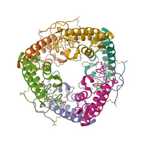
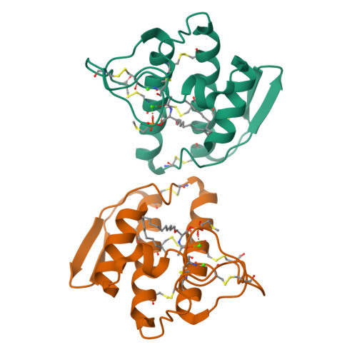
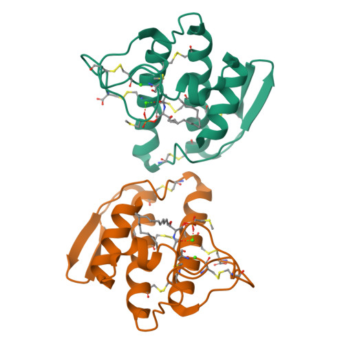
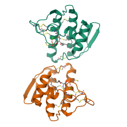
    It is often found in the crystallization of enzyme-inhibitor complexes that an inhibitor causes crystal packing which is different to that of native protein. This is the case for crystals of human non-pancreatic secreted phospholipase A(2) (124 residues) containing six molecules in the asymmetric unit when the protein is complexed with a potential acylamino analogue of a phospholid. The hexameric structure was determined by molecular replacement using the structure of monomeric native protein as a probe. As an extension to the experiment, it was tested whether a backbone polypeptide composed of 17% of a known monomeric structure could find its correct position on a target molecule in molecular replacement. A probe model composed of the backbone atoms of the N-terminal 77 residues of lysine-, arginine-, ornithine-binding protein (LAO, a total of 238 residues) liganded with lysine correctly finds its position on LAO liganded with histidine which crystallizes as a monomer in the asymmetric unit. The results indicate that as little as 17% of total diffracting matter can be used in molecular replacement to solve crystal structures or to obtain phase information which can be combined with phases obtained by the isomorphous-replacement method.


  • Organizational Affiliation

    Department of Macromolecular Science, SmithKline Beecham Pharmaceuticals, King of Prussia, PA 19406, USA.


Macromolecules
Find similar proteins by:  (by identity cutoff)  |  3D Structure
Entity ID: 1
MoleculeChains Sequence LengthOrganismDetailsImage
PHOSPHOLIPASE A2
A, B, C, D, E
A, B, C, D, E, F
124Homo sapiensMutation(s): 0 
EC: 3.1.1.4
UniProt & NIH Common Fund Data Resources
Find proteins for P14555 (Homo sapiens)
Explore P14555 
Go to UniProtKB:  P14555
PHAROS:  P14555
GTEx:  ENSG00000188257 
Entity Groups  
Sequence Clusters30% Identity50% Identity70% Identity90% Identity95% Identity100% Identity
UniProt GroupP14555
Sequence Annotations
Expand
  • Reference Sequence
Small Molecules
Ligands 2 Unique
IDChains Name / Formula / InChI Key2D Diagram3D Interactions
INB
Query on INB

Download Ideal Coordinates CCD File 
I [auth A]
L [auth B]
O [auth C]
R [auth D]
U [auth E]
I [auth A],
L [auth B],
O [auth C],
R [auth D],
U [auth E],
X [auth F]
1-OCTADECYL-2-ACETAMIDO-2-DEOXY-SN-GLYCEROL-3-PHOSPHOETHYLMETHYL SULFIDE
C26 H54 N O6 P S
ZULTVWFLRZJENJ-AREMUKBSSA-N
CA
Query on CA

Download Ideal Coordinates CCD File 
G [auth A]
H [auth A]
J [auth B]
K [auth B]
M [auth C]
G [auth A],
H [auth A],
J [auth B],
K [auth B],
M [auth C],
N [auth C],
P [auth D],
Q [auth D],
S [auth E],
T [auth E],
V [auth F],
W [auth F]
CALCIUM ION
Ca
BHPQYMZQTOCNFJ-UHFFFAOYSA-N
Experimental Data & Validation

Experimental Data

  • Method: X-RAY DIFFRACTION
  • Resolution: 2.57 Å
  • R-Value Work: 0.214 
  • R-Value Observed: 0.214 
  • Space Group: P 1 21 1
Unit Cell:
Length ( Å )Angle ( ˚ )
a = 64.51α = 90
b = 114.72β = 120.13
c = 64.43γ = 90
Software Package:
Software NamePurpose
X-PLORmodel building
X-PLORrefinement
X-PLORphasing

Structure Validation

View Full Validation Report


Ligand Structure Quality Assessment 


Entry History 

Deposition Data

  • Released Date: 1995-07-31 
  • Deposition Author(s): Oh, B.-H.

Revision History  (Full details and data files)

  • Version 1.0: 1995-07-31
    Type: Initial release
  • Version 1.1: 2008-03-24
    Changes: Version format compliance
  • Version 1.2: 2011-07-13
    Changes: Version format compliance
  • Version 1.3: 2024-06-05
    Changes: Data collection, Database references, Derived calculations, Other
  • Version 1.4: 2024-10-16
    Changes: Structure summary