Crystal structures of Paracoccus denitrificans aromatic amino acid aminotransferase: a substrate recognition site constructed by rearrangement of hydrogen bond network.
Okamoto, A., Nakai, Y., Hayashi, H., Hirotsu, K., Kagamiyama, H.(1998) J Mol Biology 280: 443-461
- PubMed: 9665848 Search on PubMed
- DOI: https://doi.org/10.1006/jmbi.1998.1869
- Primary Citation Related Structures:
1AY4, 1AY5, 1AY8 - PubMed Abstract:
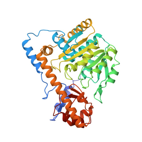
Aminotransferase reversibly catalyzes the transamination reaction by a ping-pong bi-bi mechanism with pyridoxal 5'-phosphate (PLP) as a cofactor. Various kinds of aminotransferases developing into catalysts for particular substrates have been reported. Among the aminotransferases, aromatic amino acid aminotransferase (EC 2.6.1. 57) catalyzes the transamination reaction with both acidic substrates and aromatic substrates. To elucidate the multiple substrate recognition mechanism, we determined the crystal structures of aromatic amino acid aminotransferase from Paracoccus denitrificans (pdAroAT): unliganded pdAroAT, pdAroAT in a complex with maleate as an acidic substrate analog, and pdAroAT in a complex with 3-phenylpropionate as an aromatic substrate analog at 2.33 A, 2. 50 A and 2.30 A resolution, respectively. The pdAroAT molecule is a homo-dimer. Each subunit has 394 amino acids and one PLP and is divided into small and large domains. The overall structure of pdAroAT is essentially identical to that of aspartate aminotransferase (AspAT) which catalyzes the transamination reaction with only an acidic amino acid. On binding the acidic substrate analog, arginine 292 and 386 form end-on salt bridges with carboxylates of the analog. Furthermore, binding of the substrate induces the domain movement to close the active site. The recognition mechanism for the acidic substrate analog in pdAroAT is identical to that observed in AspAT. Binding of the aromatic substrate analog causes reorientation of the side-chain of the residues, lysine 16, asparagine 142, arginine 292* and serine 296*, and changes in the position of water molecules in the active site to form a new hydrogen bond network in contrast to the active site structure of pdAroAT in the complex with an acidic substrate analog. Consequently, the rearrangement of the hydrogen bond network can form recognition sites for both acidic and aromatic side-chains of the substrate without a conformational change in the backbone structure in pdAroAT.
- Department of Biochemistry, Osaka Medical College, Takatsuki, Osaka, 569-8686, Japan.
Organizational Affiliation: