Crystal structures of the copper-containing amine oxidase from Arthrobacter globiformis in the holo and apo forms: implications for the biogenesis of topaquinone.
Wilce, M.C., Dooley, D.M., Freeman, H.C., Guss, J.M., Matsunami, H., McIntire, W.S., Ruggiero, C.E., Tanizawa, K., Yamaguchi, H.(1997) Biochemistry 36: 16116-16133
- PubMed: 9405045
- DOI: https://doi.org/10.1021/bi971797i
- Primary Citation of Related Structures:
1AV4, 1AVK, 1AVL - PubMed Abstract:
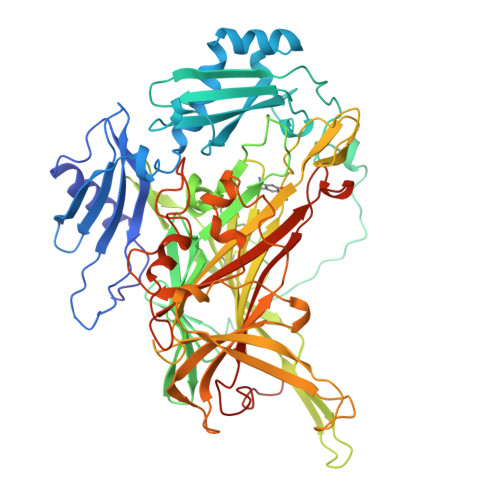
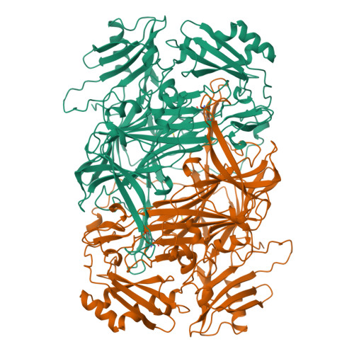
The crystal structures of the copper enzyme phenylethylamine oxidase from the Gram-positive bacterium Arthrobacter globiformis (AGAO) have been determined and refined for three forms of the enzyme: the holoenzyme in its active form (at 2.2 A resolution), the holoenzyme in an inactive form (at 2.8 A resolution), and the apoenzyme (at 2.2 A resolution). The holoenzyme has a topaquinone (TPQ) cofactor formed from the apoenzyme by the post-translational modification of a tyrosine residue in the presence of Cu2+. Significant differences between the three forms of AGAO are limited to the active site. The polypeptide fold is closely similar to those of the amine oxidases from Escherichia coli [Parsons, M. R., et al. (1995) Structure 3, 1171-1184] and pea seedlings [Kumar, V., et al. (1996) Structure 4, 943-955]. In the active form of holo-AGAO, the active-site Cu atom is coordinated by three His residues and two water molecules in an approximately square-pyramidal arrangement. In the inactive form, the Cu atom is coordinated by the same three His residues and by the phenolic oxygen of the TPQ, the geometry being quasi-trigonal-pyramidal. There is evidence of disorder in the crystals of both forms of holo-AGAO. As a result, only the position of the aromatic group of the TPQ cofactor, but not its orientation about the Cbeta-Cgamma bond, is determined unequivocally. In apo-AGAO, electron density consistent with an unmodified Tyr occurs at a position close to that of the TPQ in the inactive holo-AGAO. This observation has implications for the biogenesis of TPQ. Two features which have not been described previously in amine oxidase structures are a channel from the molecular surface to the active site and a solvent-filled cavity at the major interface between the two subunits of the dimer.
Organizational Affiliation:
School of Chemistry and Department of Biochemistry, University of Sydney, New South Wales 2006, Australia.


