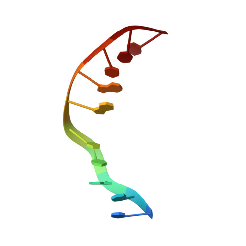
Refined solution structure of 8,9-dihydro-8-(N7-guanyl)-9-hydroxyaflatoxin B1 opposite CpA in the complementary strand of an oligodeoxynucleotide duplex as determined by 1H NMR.
Johnston, D.S., Stone, M.P.(1995) Biochemistry 34: 14037-14050
- PubMed: 7578001
- DOI: https://doi.org/10.1021/bi00043a009
- Primary Citation of Related Structures:
1AG5 - PubMed Abstract:
The solution structure of d(CCATCAFBGATCC).d(GGATCAGATGG), containing the 8,9-dihydro-8-(N7-guanyl)-9-hydroxyaflatoxin B1 adduct, was refined using molecular dynamics restrained by NOE data obtained from 1H NMR. The modified guanosine was positioned opposite cytosine, while the aflatoxin moiety was positioned opposite adenosine in the complementary strand. Sequential 1H NOEs were interrupted between C5 and AFBG6, but intrastrand NOEs were traced through the aflatoxin moiety, via H6a of aflatoxin and H8 of the modified guanine. Opposite the lesion, the NOE between A16 H1' and G17 H8 was weak. A total of 43 NOEs were observed between DNA protons and aflatoxin protons. Molecular dynamics calculations restrained with 259 experimental and empirical distances, and using sp2 hybridization at AFBG6 N7, refined structures with pairwise rms differences < 0.85 A, excluding terminal base pairs. Relaxation matrix calculations yielded a sixth root rms difference between refined structures and NOE intensity data of 7.3 x 10(-2). The aflatoxin moiety intercalated on the 5'-face of the modified guanine. The extra adenine A16 was inserted between base pair AFBG6.C15 and the aflatoxin moiety. A 36 degree bending between the plane of base pair AFBG6.C15 and the plane of the aflatoxin moiety was predicted. The aflatoxin moiety stacked below the top domain of the oligodeoxynucleotide, which consisted of base pairs C1.G21, C2.G20, A3.T19, T4.A18, and C5.G17. The bottom domain consisted of base pairs AFBG6.C15, A7.T14, T8.A13, C9.G12, and C10.G11. The average winding angle between base pair C5.G17, the intercalated aflatoxin moiety, A16, and base pair AFBG6.C15 was reduced to 10 degrees. The preponderance of base pair substitutions in the aflatoxin B1 mutational spectrum, particularly G-->T transversions, suggests that the stability of this modified oligodeoxynucleotide, which models a templated +1 addition mutation, does not reliably predict the frequency of frame shifts.
Organizational Affiliation:
Center in Molecular Toxicology, Vanderbilt University, Nashville, Tennessee 37235, USA.


