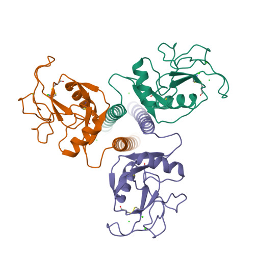
1AFD

STRUCTURAL BASIS OF GALACTOSE RECOGNITION IN C-TYPE ANIMAL LECTINS


Experimental Data Snapshot

  • Method: X-RAY DIFFRACTION
  • Resolution: 2.00 Å
  • R-Value Free: 0.282 
  • R-Value Work: 0.234 
  • R-Value Observed: 0.234 

wwPDB Validation   3D Report Full Report


This is version 1.4 of the entry. See complete history


Literature

Structural basis of galactose recognition by C-type animal lectins.

Kolatkar, A.R.Weis, W.I.

(1996) J Biol Chem 271: 6679-6685

  • Primary Citation of Related Structures:  
    1AFA, 1AFB, 1AFD

  • PubMed Abstract: 

    The asialoglycoprotein receptors and many other C-type (Ca2+-dependent) animal lectins specifically recognize galactose- or N-acetylgalactosamine-terminated oligosaccharides. Analogous binding specificity can be engineered into the homologous rat mannose-binding protein A by changing three amino acids and inserting a glycine-rich loop (Iobst, S. T., and Drickamer, K. (1994) J. Biol. Chem. 269, 15512-15519). Crystal structures of this mutant complexed with beta-methyl galactoside and N-acetylgalactosamine (GalNAc) reveal that as with wild-type mannose-binding proteins, the 3- and 4-OH groups of the sugar directly coordinate Ca2+ and form hydrogen bonds with amino acids that also serve as Ca2+ ligands. The different stereochemistry of the 3- and 4-OH groups in mannose and galactose, combined with a fixed Ca2+ coordination geometry, leads to different pyranose ring locations in the two cases. The glycine-rich loop provides selectivity against mannose by holding a critical tryptophan in a position optimal for packing with the apolar face of galactose but incompatible with mannose binding. The 2-acetamido substituent of GalNAc is in the vicinity of amino acid positions identified by site-directed mutagenesis (Iobst, S. T., and Drickamer, K. (1996) J. Biol. Chem. 271, 6686-6693) as being important for the formation of a GalNAc-selective binding site.


  • Organizational Affiliation

    Department of Structural Biology, Stanford University School of Medicine, Stanford, California 94305, USA.


Macromolecules
Find similar proteins by:  (by identity cutoff)  |  3D Structure
Entity ID: 1
MoleculeChains Sequence LengthOrganismDetailsImage
MANNOSE-BINDING PROTEIN-AA [auth 1],
B [auth 2],
C [auth 3]
154Rattus norvegicusMutation(s): 5 
UniProt
Find proteins for P19999 (Rattus norvegicus)
Explore P19999 
Go to UniProtKB:  P19999
Entity Groups  
Sequence Clusters30% Identity50% Identity70% Identity90% Identity95% Identity100% Identity
UniProt GroupP19999
Sequence Annotations
Expand
  • Reference Sequence
Small Molecules
Ligands 2 Unique
IDChains Name / Formula / InChI Key2D Diagram3D Interactions
CA
Query on CA

Download Ideal Coordinates CCD File 
D [auth 1]
E [auth 1]
F [auth 1]
H [auth 2]
I [auth 2]
D [auth 1],
E [auth 1],
F [auth 1],
H [auth 2],
I [auth 2],
J [auth 3],
K [auth 3],
L [auth 3]
CALCIUM ION
Ca
BHPQYMZQTOCNFJ-UHFFFAOYSA-N
CL
Query on CL

Download Ideal Coordinates CCD File 
G [auth 1],
M [auth 3]
CHLORIDE ION
Cl
VEXZGXHMUGYJMC-UHFFFAOYSA-M
Experimental Data & Validation

Experimental Data

  • Method: X-RAY DIFFRACTION
  • Resolution: 2.00 Å
  • R-Value Free: 0.282 
  • R-Value Work: 0.234 
  • R-Value Observed: 0.234 
  • Space Group: C 1 2 1
Unit Cell:
Length ( Å )Angle ( ˚ )
a = 80.4α = 90
b = 84.7β = 105.4
c = 98γ = 90
Software Package:
Software NamePurpose
DENZOdata reduction
SCALEPACKdata scaling
X-PLORmodel building
X-PLORrefinement
X-PLORphasing

Structure Validation

View Full Validation Report


Entry History 

Deposition Data

Revision History  (Full details and data files)

  • Version 1.0: 1996-04-03
    Type: Initial release
  • Version 1.1: 2008-03-03
    Changes: Version format compliance
  • Version 1.2: 2011-07-13
    Changes: Version format compliance
  • Version 1.3: 2021-11-03
    Changes: Database references, Derived calculations
  • Version 1.4: 2024-11-20
    Changes: Data collection, Structure summary