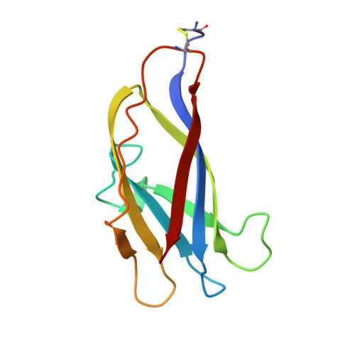
Solution structure of the granular starch binding domain of Aspergillus niger glucoamylase bound to beta-cyclodextrin.
Sorimachi, K., Le Gal-Coeffet, M.F., Williamson, G., Archer, D.B., Williamson, M.P.(1997) Structure 5: 647-661
- PubMed: 9195884 Search on PubMed
- DOI: https://doi.org/10.1016/s0969-2126(97)00220-7
- Primary Citation Related Structures:
1AC0, 1ACZ - PubMed Abstract:
Carbohydrate-binding domains are usually small and physically separate from the catalytic domains of hydrolytic enzymes. Glucoamylase 1 (G1) from Aspergillus niger, an enzyme used widely in the food and brewing industries, contains a granular starch binding domain (SBD) which is separated from the catalytic domain by a semi-rigid linker. The aim of this study was to determine how the SBD binds to starch, and thereby more generally to throw light on the role of carbohydrate-binding domains in the hydrolysis of insoluble polysaccharides. The solution structure of the SBD of A. niger G1 bound to beta-cyclodextrin (betaCD), a cyclic starch analogue, shows that the well-defined beta-sheet structure seen in the free SBD is maintained in the SBD-betaCD complex. The main differences between the free and bound states of the SBD are observed in loop regions, in or near the two starch-binding sites. The two binding sites, each of which binds one molecule of betaCD, are structurally different. Binding site 1 is small and accessible, and its structure changes very little upon ligand binding. Site 2 is longer and undergoes a significant structural change on binding. Part of this site comprises a flexible loop, which appears to allow the SBD to bind to starch strands in a range of orientations. The two starch-binding sites of the SBD probably differ functionally as well as structurally; site 1 probably acts as the initial starch recognition site, whereas site 2 is involved in specific recognition of appropriate regions of starch. The two starch strands are bound at approximately 90 degrees to each other. This may be functionally important, as it may force starch strands apart thus increasing the hydrolyzable surface, or alternatively it may localize the enzyme to noncrystalline (more hydrolyzable) areas of starch. The region of the SBD where the linker to the catalytic domain is attached is flexible, allowing the catalytic site to access a large surface area of the starch granules.
- Krebs Institute for Biomolecular Research Department of Molecular Biology and Biotechnology University of Sheffield Firth Court, Western Bank, Sheffield, S10 2TN, UK.
Organizational Affiliation: