1A4B | pdb_00001a4b

AZURIN MUTANT WITH MET 121 REPLACED BY HIS, PH 6.5 CRYSTAL FORM, DATA COLLECTED AT-180 DEGREES CELSIUS


Experimental Data Snapshot

  • Method: X-RAY DIFFRACTION
  • Resolution: 1.91 Å
  • R-Value Work: 
    0.202 (Depositor) 
  • R-Value Observed: 
    0.202 (Depositor) 

Starting Model: experimental
View more details

wwPDB Validation 3D Report Full Report

Validation slider image for 1A4B

This is version 2.2 of the entry. See complete history

Literature

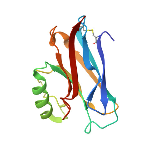
Rack-induced metal binding vs. flexibility: Met121His azurin crystal structures at different pH.

Messerschmidt, A.Prade, L.Kroes, S.J.Sanders-Loehr, J.Huber, R.Canters, G.W.

(1998) Proc Natl Acad Sci U S A 95: 3443-3448

  • DOI: https://doi.org/10.1073/pnas.95.7.3443
  • Primary Citation Related Structures: 
    1A4A, 1A4B, 1A4C

  • PubMed Abstract: 

    The rack-induced bonding mechanism of metals to proteins is a useful concept for explaining the generation of metal sites in electron transfer proteins, such as the blue copper proteins, that are designed for rapid electron transfer. The trigonal pyramidal structure imposed by the protein with three strong equatorial ligands (one Cys and two His) provides a favorable geometry for both cuprous and cupric oxidation states. However, the crystal structures of the Met121His mutant of azurin from Alcaligenes denitrificans at pH 6.5 (1.89- and 1.91-A resolutions) and pH 3.5 (2.45-A resolution) show that the preformed metal binding cavity in the protein is more flexible than expected. At high pH (6.5), the Cu site retains the same three equatorial ligands as in the wild-type azurin and adds His121 as a fourth strong ligand, creating a tetrahedral copper site geometry with a green color referred to as 1.5 type. In the low pH (3.5) structure, the protonation of His121 causes a conformational change in residues 117-123, moving His121 away from the copper. The empty coordination site is occupied by an oxygen atom of a nitrate molecule of the buffer solution. This axial ligand is coordinated less strongly, generating a distorted tetrahedral copper geometry with a blue color and spectroscopic properties of a type-1 site. These crystal structures demonstrate that blue copper proteins are flexible enough to permit a range of movement of the Cu atom along the axial direction of the trigonal pyramid.


  • Organizational Affiliation
    • Max-Planck-Institut für Biochemie, D-82152 Martinsried, Germany. messersc@biochem.mpg.de

Macromolecule Content 

  • Total Structure Weight: 28.33 kDa 
  • Atom Count: 2,323 
  • Modeled Residue Count: 258 
  • Deposited Residue Count: 258 
  • Unique protein chains: 1

Macromolecules

Find similar proteins by:|  3D Structure
Entity ID: 1
MoleculeChains  Sequence LengthOrganismDetailsImage
AZURIN
A, B
129Achromobacter denitrificansMutation(s): 1 
Gene Names: AZU
UniProt
Find proteins for P00280 (Achromobacter denitrificans)
Explore P00280 
Go to UniProtKB:  P00280
Entity Groups
Sequence Clusters30% Identity50% Identity70% Identity90% Identity95% Identity100% Identity
UniProt GroupP00280
Sequence Annotations
Expand
Reference Sequence

Experimental Data & Validation

Experimental Data

  • Method: X-RAY DIFFRACTION
  • Resolution: 1.91 Å
  • R-Value Work:  0.202 (Depositor) 
  • R-Value Observed: 0.202 (Depositor) 
Space Group: C 2 2 21
Unit Cell:
Length ( Å )Angle ( ˚ )
a = 74.56α = 90
b = 73.57β = 90
c = 97.63γ = 90
Software Package:
Software NamePurpose
MOSFLMdata reduction
ROTAVATAdata reduction
Agrovatadata reduction
AMoREphasing
X-PLORrefinement
CCP4data scaling
ROTAVATAdata scaling

Structure Validation

View Full Validation Report


Entry History 

Deposition Data

Revision History  (Full details and data files)

  • Version 1.0: 1998-04-29
    Type: Initial release
  • Version 1.1: 2008-03-03
    Changes: Version format compliance
  • Version 1.2: 2011-07-13
    Changes: Version format compliance
  • Version 1.3: 2017-11-29
    Changes: Refinement description
  • Version 2.0: 2021-11-03
    Changes: Atomic model, Database references, Derived calculations
  • Version 2.1: 2023-08-02
    Changes: Advisory, Refinement description
  • Version 2.2: 2024-11-06
    Changes: Data collection, Structure summary