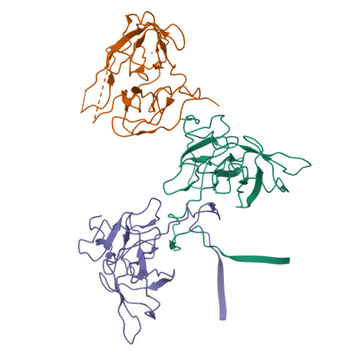
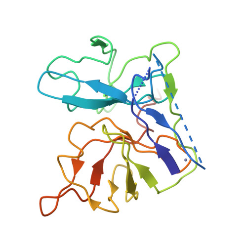
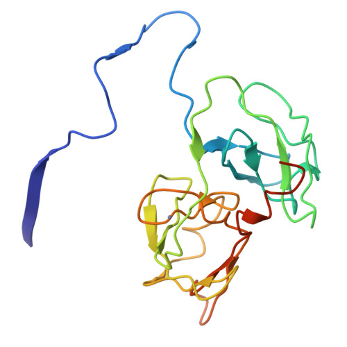
1A1Q

HEPATITIS C VIRUS NS3 PROTEINASE


Experimental Data Snapshot

  • Method: X-RAY DIFFRACTION
  • Resolution: 2.40 Å
  • R-Value Free: 0.320 
  • R-Value Work: 0.225 
  • R-Value Observed: 0.225 

wwPDB Validation   3D Report Full Report


This is version 1.3 of the entry. See complete history


Literature

The crystal structure of hepatitis C virus NS3 proteinase reveals a trypsin-like fold and a structural zinc binding site.

Love, R.A.Parge, H.E.Wickersham, J.A.Hostomsky, Z.Habuka, N.Moomaw, E.W.Adachi, T.Hostomska, Z.

(1996) Cell 87: 331-342

  • DOI: https://doi.org/10.1016/s0092-8674(00)81350-1
  • Primary Citation of Related Structures:  
    1A1Q

  • PubMed Abstract: 

    During replication of hepatitis C virus (HCV), the final steps of polyprotein processing are performed by a viral proteinase located in the N-terminal one-third of nonstructural protein 3. The structure of NS3 proteinase from HCV BK strain was determined by X-ray crystallography at 2.4 angstrom resolution. NS3P folds as a trypsin-like proteinase with two beta barrels and a catalytic triad of His-57, Asp-81, Ser-139. The structure has a substrate-binding site consistent with the cleavage specificity of the enzyme. Novel features include a structural zinc-binding site and a long N-terminus that interacts with neighboring molecules by binding to a hydrophobic surface patch.


  • Organizational Affiliation

    Agouron Pharmaceuticals, Inc., San Diego, California 92121, USA.


Macromolecules
Find similar proteins by:  (by identity cutoff)  |  3D Structure
Entity ID: 1
MoleculeChains Sequence LengthOrganismDetailsImage
NS3 PROTEINASE
A, B, C
189Hepacivirus hominisMutation(s): 0 
Gene Names: CDNA
UniProt
Find proteins for P26663 (Hepatitis C virus genotype 1b (isolate BK))
Explore P26663 
Go to UniProtKB:  P26663
Entity Groups  
Sequence Clusters30% Identity50% Identity70% Identity90% Identity95% Identity100% Identity
UniProt GroupP26663
Sequence Annotations
Expand
  • Reference Sequence
Experimental Data & Validation

Experimental Data

  • Method: X-RAY DIFFRACTION
  • Resolution: 2.40 Å
  • R-Value Free: 0.320 
  • R-Value Work: 0.225 
  • R-Value Observed: 0.225 
  • Space Group: H 3 2
Unit Cell:
Length ( Å )Angle ( ˚ )
a = 133α = 90
b = 133β = 90
c = 223γ = 120
Software Package:
Software NamePurpose
PHASESphasing
X-PLORmodel building
X-PLORrefinement
DENZOdata reduction
SCALEPACKdata scaling
X-PLORphasing

Structure Validation

View Full Validation Report


Entry History 

Deposition Data

Revision History  (Full details and data files)

  • Version 1.0: 1998-03-25
    Type: Initial release
  • Version 1.1: 2008-03-24
    Changes: Version format compliance
  • Version 1.2: 2011-07-13
    Changes: Version format compliance
  • Version 1.3: 2024-02-07
    Changes: Data collection, Database references, Derived calculations