13HE | pdb_000013he

CryoEM structure of AdhE Spirosome from Clostridium thermocellum uncovered by visual proteomics.

  • Classification: PROTEIN FIBRIL
  • Organism(s): Acetivibrio thermocellus DSM 1313
  • Mutation(s): No 

  • Deposited: 2026-05-06 Released: 2026-06-24 
  • Deposition Author(s): Agdanowski, M.P., Rodriguez, J.A.
  • Funding Organization(s): Department of Energy (DOE, United States), National Science Foundation (NSF, United States), National Institutes of Health/National Institute of General Medical Sciences (NIH/NIGMS)

Experimental Data Snapshot

  • Method: ELECTRON MICROSCOPY
  • Resolution: 4.07 Å
  • Aggregation State: FILAMENT 
  • Reconstruction Method: HELICAL 

Starting Model: experimental
View more details

wwPDB Validation 3D Report Full Report

Validation slider image for 13HE

This is version 2.0 of the entry. See complete history. 

Literature

Visual exoproteomics of Clostridium thermocellum during anaerobic biomass-degradation identifies functional spirosomes.

Agdanowski, M.P., Kensil, M.J., Moser, T.H., Humm, E., Guandique, Y.I., Mason-Chalmers, K., Pia, V.G., Al-Set, T., Ogorzalek Loo, R.R., Evans, J.E., Gunsalus, R.P., Loo, J.A., Rodriguez, J.A.

(2026) bioRxiv 

  • DOI: https://doi.org/10.64898/2026.05.21.726627
  • Primary Citation Related Structures: 
    13HE

  • PubMed Abstract: 

    Visual proteomics enables the study of low-abundance proteins and identification of unknown complexes from heterogeneous samples by complementing high-resolution cryogenic electron microscopy (cryoEM) with external inputs on protein identity such as mass spectrometry. Using this approach, we interrogated the exoproteome of the anaerobic cellulose-degrading bacterium Clostridium thermocellum as it carried out biomass degradation. Mass spectrometry indicated a broad exoproteome composition, including cellulose degrading machinery CelA and CipA. A focus on large exoproteome assemblies revealed abundant protein filaments and pleomorphic vesicular structures. Analysis of the most abundant protein filaments yielded an ~4 resolution native structure that, aided by mass spectrometry, de novo modeling, and structural searching, was found to be the aldehyde-alcohol dehydrogenase (AdhE) spirosome. AdhE contained both NAD + and Fe in their expected binding sites and biochemical and structural analyses of enriched spirosome preparations indicated they were functional. Altered NADH solution concentrations triggered conformational changes in the exoproteomic spirosomes, and the constituent AdhE remained capable of ethanol production. Although the basis for functional extracellular spirosome accumulation in live anaerobic C. thermocellum cultures remains unclear, their abundance in crude exoproteomes suggests their presence could influence biomass fueled C. thermocellum growth.


  • Organizational Affiliation: 
    • Department of Chemistry and Biochemistry; UCLA-DOE Institute for Genomics and Proteomics; University of California, Los Angeles (UCLA), Los Angeles, CA 90095, USA.

Macromolecule Content 

  • Total Structure Weight: 573.04 kDa 
  • Atom Count: 40,128 
  • Modeled Residue Count: 5,166 
  • Deposited Residue Count: 5,166 
  • Unique protein chains: 1

Macromolecules

Find similar proteins by:|  3D Structure
Entity ID: 1
MoleculeChains  Sequence LengthOrganismDetailsImage
Aldehyde-alcohol dehydrogenase
A, B, C, D, E
A, B, C, D, E, F
861Acetivibrio thermocellus DSM 1313Mutation(s): 0 
UniProt
Find proteins for A0A0H3W5U9 (Acetivibrio thermocellus)
Explore A0A0H3W5U9 
Go to UniProtKB:  A0A0H3W5U9
Entity Groups
Sequence Clusters30% Identity50% Identity70% Identity90% Identity95% Identity100% Identity
UniProt GroupA0A0H3W5U9
Sequence Annotations
Expand
Reference Sequence

Small Molecules

Ligands 2 Unique
IDChains Name / Formula / InChI Key2D Diagram3D Interactions
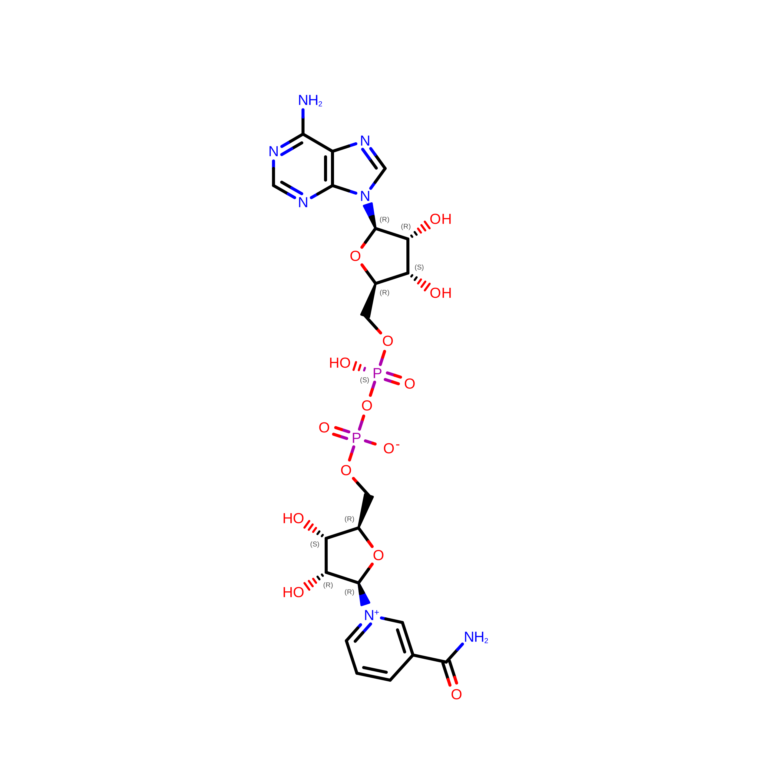
NAD

Query on NAD


Download:Ideal Coordinates CCD File
H [auth A]
J [auth B]
L [auth C]
N [auth D]
P [auth E]
H [auth A],
J [auth B],
L [auth C],
N [auth D],
P [auth E],
R [auth F]
NICOTINAMIDE-ADENINE-DINUCLEOTIDE
C21 H27 N7 O14 P2
BAWFJGJZGIEFAR-NNYOXOHSSA-N
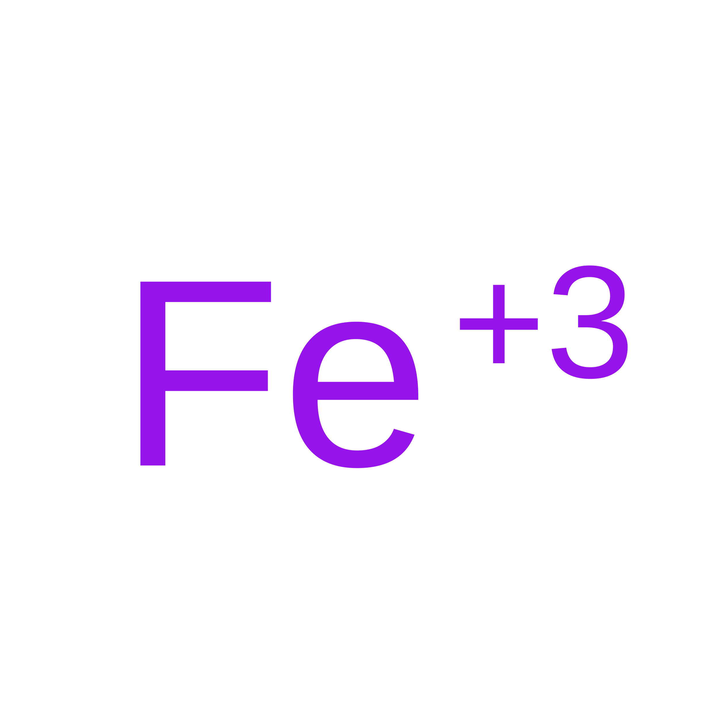
FE

Query on FE


Download:Ideal Coordinates CCD File
G [auth A]
I [auth B]
K [auth C]
M [auth D]
O [auth E]
G [auth A],
I [auth B],
K [auth C],
M [auth D],
O [auth E],
Q [auth F]
FE (III) ION
Fe
VTLYFUHAOXGGBS-UHFFFAOYSA-N

Experimental Data & Validation

Experimental Data

  • Method: ELECTRON MICROSCOPY
  • Resolution: 4.07 Å
  • Aggregation State: FILAMENT 
  • Reconstruction Method: HELICAL 
EM Software:
TaskSoftware PackageVersion
RECONSTRUCTIONcryoSPARC4.7.0
MODEL REFINEMENTPHENIX2.0_5936

Structure Validation

View Full Validation Report


Entry History 

& Funding Information

Deposition Data


Funding OrganizationLocationGrant Number
Department of Energy (DOE, United States)United StatesDE-FC02-02ER63421
National Science Foundation (NSF, United States)United StatesDMR-1548924
National Institutes of Health/National Institute of General Medical Sciences (NIH/NIGMS)United StatesR35GM128867
National Institutes of Health/National Institute of General Medical Sciences (NIH/NIGMS)United StatesR35GM145286
Department of Energy (DOE, United States)United StatesKP1607011

Revision History  (Full details and data files)

  • Version 1.0: 2026-06-24
    Type: Initial release
  • Version 2.0: 2026-09-16
    Type: Coordinate replacement
    Reason: Model orientation/position
    Changes: Atomic model, Author supporting evidence, Data collection, Data processing, Database references, Derived calculations, Refinement description, Structure summary