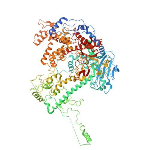
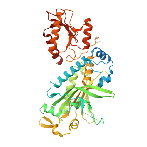
12SL | pdb_000012sl

Structure of a Strand-Displacement Complex of Human Mitochondrial DNA Polymerase Gamma


Experimental Data Snapshot

  • Method: ELECTRON MICROSCOPY
  • Resolution: 2.93 Å
  • Aggregation State: PARTICLE 
  • Reconstruction Method: SINGLE PARTICLE 

Starting Model: experimental
View more details

wwPDB Validation 3D Report Full Report

Validation slider image for 12SL

This is version 1.0 of the entry. See complete history. 

Literature

Molecular and structural basis for replication initiation and strand separation by human mitochondrial DNA polymerase gamma.

Sokolova, V., Buchel, G., Strock, S., Nayak, A.R., Temiakov, D.

(2026) Nucleic Acids Res 54

  • DOI: https://doi.org/10.1093/nar/gkag925
  • Primary Citation Related Structures: 
    12SL, 9PCK

  • PubMed Abstract: 

    Defects in human mitochondrial DNA (mtDNA) replication can lead to somatic mutations associated with a range of devastating mitochondrial diseases. However, the molecular mechanisms governing the earliest steps of mtDNA replication and their fidelity remain poorly understood. Here, we found that DNA polymerase gamma (Polγ) forms stable complexes with RNA-DNA primer-template substrates, exhibiting greater stability and lower misincorporation than on DNA-primed substrates. Structural analysis revealed that Polγ interacts with the 2'-OH groups of ribose within the first four nucleotides of the primer, explaining the stability of complexes that utilize RNA primers. Although Polγ requires TWINKLE to extend RNA primers, its intrinsic strand-displacement activity allows it to extend DNA primers independently. Structural data further show that the strand-separation mechanism in human Polγ is distinct from that of its yeast paralog, Mip1, and involves previously unresolved elements-the catcher and a GP loop in the exonuclease domain-that support intrinsic strand-displacement synthesis by Polγ. Structure-guided mutagenesis of elements involved in strand separation supports these structural observations. Together, our study provides mechanistic insight into mtDNA replication initiation and strand separation and has implications for understanding the molecular basis of mitochondrial disease.


  • Organizational Affiliation: 
    • Department of Biochemistry and Molecular Biology, Thomas Jefferson University; 1020 Locust St, Philadelphia, PA 19107, United States.

Macromolecule Content 

  • Total Structure Weight: 263.71 kDa 
  • Atom Count: 14,769 
  • Modeled Residue Count: 1,825 
  • Deposited Residue Count: 2,252 
  • Unique protein chains: 2
  • Unique nucleic acid chains: 2

Macromolecules


Find similar proteins by:|  3D Structure
Entity ID: 1
MoleculeChains  Sequence LengthOrganismDetailsImage
DNA polymerase subunit gamma-11,239Homo sapiensMutation(s): 0 
Gene Names: POLG, MDP1, POLG1, POLGA
EC: 2.7.7.7 (PDB Primary Data), 3.1.11 (UniProt), 4.2.99 (UniProt)
UniProt & NIH Common Fund Data Resources
Find proteins for P54098 (Homo sapiens)
Explore P54098 
Go to UniProtKB:  P54098
PHAROS:  P54098
GTEx:  ENSG00000140521 
Entity Groups
Sequence Clusters30% Identity50% Identity70% Identity90% Identity95% Identity100% Identity
UniProt GroupP54098
Sequence Annotations
Expand
Reference Sequence
Find similar proteins by:|  3D Structure
Entity ID: 2
MoleculeChains  Sequence LengthOrganismDetailsImage
DNA polymerase subunit gamma-2, mitochondrial
B, C
485Homo sapiensMutation(s): 0 
Gene Names: POLG2, MTPOLB
EC: 2.7.7.7
UniProt & NIH Common Fund Data Resources
Find proteins for Q9UHN1 (Homo sapiens)
Explore Q9UHN1 
Go to UniProtKB:  Q9UHN1
PHAROS:  Q9UHN1
GTEx:  ENSG00000256525 
Entity Groups
Sequence Clusters30% Identity50% Identity70% Identity90% Identity95% Identity100% Identity
UniProt GroupQ9UHN1
Sequence Annotations
Expand
Reference Sequence
Find similar nucleic acids by:  Sequence
Entity ID: 3
MoleculeChains LengthOrganismImage
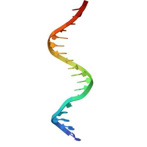
primer DNAD [auth P]19synthetic construct
Sequence Annotations
Expand
Reference Sequence
Find similar nucleic acids by:  Sequence
Entity ID: 4
MoleculeChains LengthOrganismImage
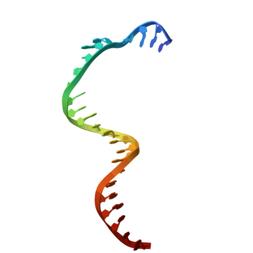
Template DNAE [auth T]24synthetic construct
Sequence Annotations
Expand
Reference Sequence

Experimental Data & Validation

Experimental Data

  • Method: ELECTRON MICROSCOPY
  • Resolution: 2.93 Å
  • Aggregation State: PARTICLE 
  • Reconstruction Method: SINGLE PARTICLE 
EM Software:
TaskSoftware PackageVersion
MODEL REFINEMENTPHENIX2.0-5936
RECONSTRUCTIONcryoSPARC4.6

Structure Validation

View Full Validation Report


Entry History 

& Funding Information

Deposition Data


Funding OrganizationLocationGrant Number
National Institutes of Health/National Institute of General Medical Sciences (NIH/NIGMS)United StatesNIH GM131832

Revision History  (Full details and data files)

  • Version 1.0: 2026-10-07
    Type: Initial release