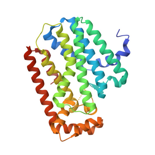
12YZ | pdb_000012yz

Crystal structure of human K293st geranylgeranyl diphosphate synthase with 5 geranylgeranyl diphosphate bound and 1 magnesium only subunits


Experimental Data Snapshot

  • Method: X-RAY DIFFRACTION
  • Resolution: 1.82 Å
  • R-Value Free: 
    0.202 (Depositor), 0.204 (DCC) 
  • R-Value Work: 
    0.169 (Depositor), 0.170 (DCC) 
  • R-Value Observed: 
    0.170 (Depositor) 

Starting Model: experimental
View more details

wwPDB Validation 3D Report Full Report

Validation slider image for 12YZ

Ligand Structure Quality Assessment 


This is version 1.0 of the entry. See complete history. 

Literature

Mapping Ligand-Induced Conformational Changes during the Enzymatic Cycle of Human Geranylgeranyl Diphosphate Synthase.

Pham, A.C., Struble, L.R., Amunugama, L., Shaw, J.B., Stachowski, T.R., Holstein, S.A., Borgstahl, G.E.O.

(2026) Biochemistry 

  • DOI: https://doi.org/10.1021/acs.biochem.6c00411
  • Primary Citation Related Structures: 
    12YZ, 12ZR

  • PubMed Abstract: 

    Geranylgeranyl diphosphate synthase (GGDPS) catalyzes the condensation reaction between two isoprenoid substrates: farnesyl diphosphate (FDP) and isopentenyl pyrophosphate (IPP), to form geranylgeranyl diphosphate (GGDP). Due to the flexible nature of the GGDPS active site and its capacity to bind multiple ligands during the reaction coordinate, understanding the structural changes that enable it to accommodate different ligands and catalyze the reaction is essential. While structures exist of substrate and product-bound GGDPS, a lack of high-resolution apo or Mg2+-only structures prevents complete understanding of the enzyme active site. Here, we report apo and Mg2+-only structures of human GGDPS, investigating the key differences between them and published GGDPS structures relating to the enzymatic cycle. A crystal structure resolved to 1.82 Å, the highest-resolution structure of human GGDPS to date, contains five product-bound subunits and one Mg2+-only subunit. The first cryo-EM structure of apo GGDPS is also reported, which exhibited an open hexamer conformation. Comparing the Mg2+-only and apo structures shows that the active site is posed and ready to bind Mg2+ without significant conformational changes. Comparing Mg2+-only to structures with either of the substrates or product bound, we observe changes in residues that actively obstruct or encourage the binding of certain ligands. We also propose an enzymatic mechanism for the structural states of GGDPS and highlight the possibility of Asp188 as the general base in the condensation reaction. These findings enhance our understanding of GGDPS ligand binding, which will improve the knowledge base for rational, structure-based design of future GGDPS inhibitors.


  • Organizational Affiliation: 
    • Department of Biochemistry and Molecular Biology, University of Nebraska Medical Center, Omaha, Nebraska68198, United States.

Macromolecule Content 

  • Total Structure Weight: 207.11 kDa 
  • Atom Count: 16,173 
  • Modeled Residue Count: 1,701 
  • Deposited Residue Count: 1,764 
  • Unique protein chains: 1

Macromolecules

Find similar proteins by:|  3D Structure
Entity ID: 1
MoleculeChains  Sequence LengthOrganismDetailsImage
Geranylgeranyl pyrophosphate synthase
A, B, C, D, E
A, B, C, D, E, F
294Homo sapiensMutation(s): 0 
Gene Names: GGPS1
EC: 2.5.1 (PDB Primary Data), 2.5.1.1 (PDB Primary Data), 2.5.1.29 (PDB Primary Data), 2.5.1.10 (PDB Primary Data)
UniProt & NIH Common Fund Data Resources
Find proteins for O95749 (Homo sapiens)
Explore O95749 
Go to UniProtKB:  O95749
PHAROS:  O95749
GTEx:  ENSG00000152904 
Entity Groups
Sequence Clusters30% Identity50% Identity70% Identity90% Identity95% Identity100% Identity
UniProt GroupO95749
Sequence Annotations
Expand
Reference Sequence

Small Molecules

Ligands 2 Unique
IDChains Name / Formula / InChI Key2D Diagram3D Interactions
GRG

Query on GRG


Download:Ideal Coordinates CCD File
I [auth A],
N [auth C],
Q [auth D],
T [auth E],
W [auth F]
GERANYLGERANYL DIPHOSPHATE
C20 H36 O7 P2
OINNEUNVOZHBOX-QIRCYJPOSA-N
MG

Query on MG


Download:Ideal Coordinates CCD File
G [auth A]
H [auth A]
J [auth B]
K [auth B]
L [auth C]
G [auth A],
H [auth A],
J [auth B],
K [auth B],
L [auth C],
M [auth C],
O [auth D],
P [auth D],
R [auth E],
S [auth E],
U [auth F],
V [auth F]
MAGNESIUM ION
Mg
JLVVSXFLKOJNIY-UHFFFAOYSA-N

Experimental Data & Validation

Experimental Data

  • Method: X-RAY DIFFRACTION
  • Resolution: 1.82 Å
  • R-Value Free:  0.202 (Depositor), 0.204 (DCC) 
  • R-Value Work:  0.169 (Depositor), 0.170 (DCC) 
  • R-Value Observed: 0.170 (Depositor) 
Space Group: P 21 21 21
Unit Cell:
Length ( Å )Angle ( ˚ )
a = 86.195α = 90
b = 135.646β = 90
c = 146.023γ = 90
Software Package:
Software NamePurpose
PHENIXrefinement
XDSdata reduction
Aimlessdata scaling
PHASERphasing

Structure Validation

View Full Validation Report


Ligand Structure Quality Assessment 


Entry History 

& Funding Information

Deposition Data


Funding OrganizationLocationGrant Number
National Institutes of Health/National Cancer Institute (NIH/NCI)United StatesCA25862
National Institutes of Health/National Cancer Institute (NIH/NCI)United StatesP30CA036727
Other governmentUnited StatesP200A210032

Revision History  (Full details and data files)

  • Version 1.0: 2026-10-28
    Type: Initial release