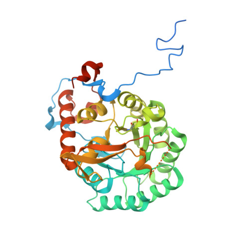
12LG | pdb_000012lg

Mtb GuaB2 dCBS in complex with inhibitor G9


Experimental Data Snapshot

  • Method: X-RAY DIFFRACTION
  • Resolution: 1.35 Å
  • R-Value Free: 
    0.175 (Depositor), 0.176 (DCC) 
  • R-Value Work: 
    0.132 (Depositor), 0.132 (DCC) 
  • R-Value Observed: 
    0.134 (Depositor) 

Starting Model: experimental
View more details

wwPDB Validation 3D Report Full Report

Validation slider image for 12LG

Ligand Structure Quality Assessment 


This is version 1.0 of the entry. See complete history. 

Literature

Development of GuaB Inhibitors to Treat Mycobacterium tuberculosis Infection.

Kofoed, E.M., Crawford, T.D., Bowman, C.M., Sims, J., Dening, T.J., Wang, S., Zhou, Y., Harris, S.F., Xu, M., Wu, P., Yang, Y., Wong, M.K., Corpuz, R., Xu, Y., Ganti, A., Ubhayakar, S., Hu, C., Clark, K.R., Cai, Z., Lai, K.W., Wang, X., Zhang, R., Koehler, M.F.T., Hanan, E.J., Aliagas, I., Ng, J., Silverman, J., Mdluli, K., Kapadia, S., Tan, M.W.

(2026) ACS Infect Dis 12: 3373-3384

  • DOI: https://doi.org/10.1021/acsinfecdis.6c00350
  • Primary Citation Related Structures: 
    12LG

  • PubMed Abstract: 

    Mycobacterium tuberculosis (Mtb) is responsible for more deaths per year than any other infectious disease. Resistance of circulating Mtb to the standard of care treatment regimen is an urgent global health crisis for which novel antibiotic treatments are needed. We have developed extremely potent, orally bioavailable, small-molecule inhibitors of inosine 5'-monophosphate dehydrogenase (GuaB) that catalyze the rate-limiting step in de novo guanine biosynthesis. The inhibitors exhibited bactericidal activity against Mtb in extracellular, intracellular, and hypoxic environments that capture features of advanced disease pathology in patients. Mtb GuaB2 inhibitors (GuaBi) were efficacious in three different in vivo mouse models of Mtb lung infection: the C57BL/6 super acute, C57BL/6 chronic, and C3HeB/FeJ (Kramnik) models. In the chronic BALB/c infection model of primarily intracellular Mtb, no benefit was observed, whereas in the C3HeB/FeJ model, a bedaquiline, pretomanid, and moxifloxacin treatment regimen that included GuaBi led to a rapid elimination of culturable Mtb infection in half of the mice. Notably, the C3HeB/FeJ model recapitulates advanced pathophysiology and heterogeneous disease that includes caseous necrotic granulomas, intracellular and extracellular reservoirs of Mtb, and lethal progressive infection. These results support further evaluation of a GuaBi-containing therapeutic regimen in relapse-capable preclinical models.


  • Organizational Affiliation: 
    • Department of Medicinal Chemistry, Genentech Inc., 1 DNA Way, South San Francisco, California94080-4990, United States.

Macromolecule Content 

  • Total Structure Weight: 44.2 kDa 
  • Atom Count: 2,769 
  • Modeled Residue Count: 338 
  • Deposited Residue Count: 422 
  • Unique protein chains: 1

Macromolecules

Find similar proteins by:|  3D Structure
Entity ID: 1
MoleculeChains  Sequence LengthOrganismDetailsImage
Inosine-5'-monophosphate dehydrogenase422Mycobacterium tuberculosisMutation(s): 0 
Gene Names: guaB, BQ2027_MB3445C
EC: 1.1.1.205
UniProt
Find proteins for P65168 (Mycobacterium bovis (strain ATCC BAA-935 / AF2122/97))
Explore P65168 
Go to UniProtKB:  P65168
Entity Groups
Sequence Clusters30% Identity50% Identity70% Identity90% Identity95% Identity100% Identity
UniProt GroupP65168
Sequence Annotations
Expand
Reference Sequence

Small Molecules

Ligands 2 Unique
IDChains Name / Formula / InChI Key2D Diagram3D Interactions
A1DCN
(Subject of Investigation/LOI)

Query on A1DCN


Download:Ideal Coordinates CCD File
C [auth A](1S,3s)-3-(4-{(1R)-1-[(1,4-dihydro-2H-pyrano[3,4-c]quinolin-9-yl)oxy]ethyl}-1H-1,2,3-triazol-1-yl)-1-methylcyclobutan-1-ol
C21 H24 N4 O3
XGRSNXCHXJRFJS-VWLHBSKXSA-N
IMP

Query on IMP


Download:Ideal Coordinates CCD File
B [auth A]INOSINIC ACID
C10 H13 N4 O8 P
GRSZFWQUAKGDAV-KQYNXXCUSA-N
Modified Residues  1 Unique
IDChains Modified ResidueModification TypeFormula2D Diagram
CSO
Query on CSO
CYSHydroxylationC3 H7 N O3 S

Experimental Data & Validation

Experimental Data

  • Method: X-RAY DIFFRACTION
  • Resolution: 1.35 Å
  • R-Value Free:  0.175 (Depositor), 0.176 (DCC) 
  • R-Value Work:  0.132 (Depositor), 0.132 (DCC) 
  • R-Value Observed: 0.134 (Depositor) 
Space Group: I 4
Unit Cell:
Length ( Å )Angle ( ˚ )
a = 88.193α = 90
b = 88.193β = 90
c = 85.037γ = 90
Software Package:
Software NamePurpose
PHENIXrefinement
Aimlessdata scaling
XDSdata reduction
PHASERphasing
PDB_EXTRACTdata extraction

Structure Validation

View Full Validation Report


Ligand Structure Quality Assessment 


Entry History 

& Funding Information

Deposition Data


Funding OrganizationLocationGrant Number
Not funded--

Revision History  (Full details and data files)

  • Version 1.0: 2026-11-04
    Type: Initial release