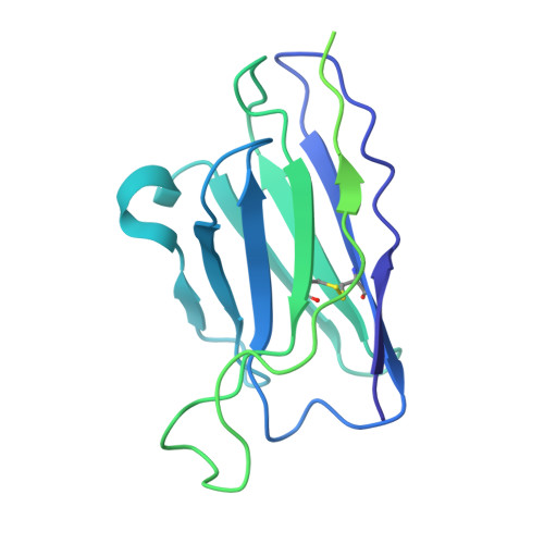
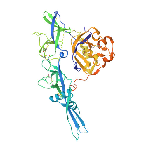
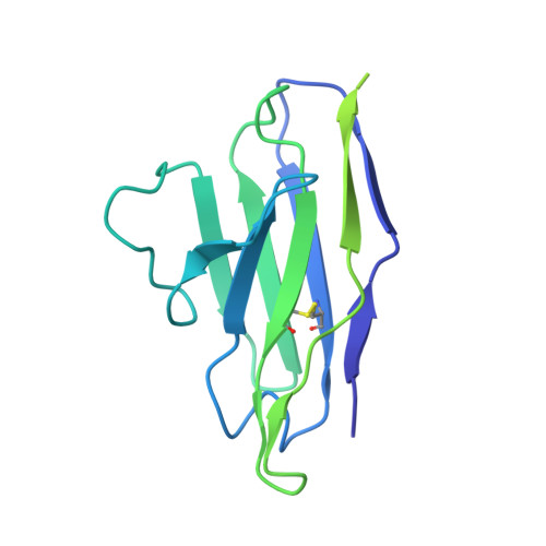
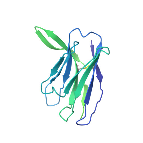
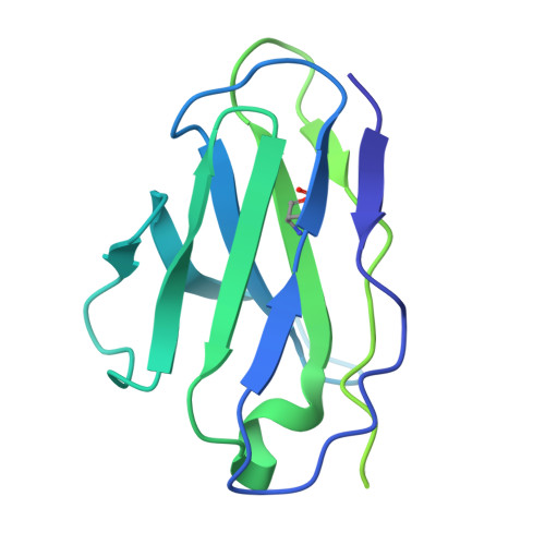
11LS | pdb_000011ls

Human Monoclonal Antibody RVC68 in complex with rabies virus glycoprotein


Experimental Data Snapshot

  • Method: ELECTRON MICROSCOPY
  • Resolution: 3.15 Å
  • Aggregation State: PARTICLE 
  • Reconstruction Method: SINGLE PARTICLE 

wwPDB Validation 3D Report Full Report

Validation slider image for 11LS

This is version 1.1 of the entry. See complete history. 

Literature

Antigenic landscape of rabies and related lyssaviruses revealed by cryo-EM.

Callaway, H.M., Zyla, D.S., Hastie, K.M., Harkins, S.S., Kothalawalage, S., Samarasinghe, N., Flynn, A., Hariharan, C., Yin, J., Corti, D., Bourhy, H., Dessain, S.K., Saphire, E.O.

(2026) Cell Rep 45: 117993-117993

  • DOI: https://doi.org/10.1016/j.celrep.2026.117993
  • Primary Citation Related Structures: 
    11IW, 11LP, 11LQ, 11LR, 11LS

  • PubMed Abstract: 

    Rabies continues to kill over 60,000 people per year despite life-saving vaccines and post-exposure treatments and costs billions of dollars in prevention and treatment. Preventing rabies deaths and reducing the global economic burden of the virus will require both developing a monoclonal antibody cocktail to replace human serum in treatment and improving rabies vaccines to elicit long-lasting protection. Here, we solve nine cryo-electron microscopy (cryo-EM) structures of neutralizing monoclonal antibodies (mAbs) in complex with the rabies virus glycoprotein (RABV-G). The nine structures span three known antigenic sites plus two additional antigenic sites, not among the five classically identified sites. We find that these antigenic sites, V and VI, are broadly cross-reactive across lyssaviruses, whereas immunodominant sites II/IV and III are rabies specific. Across the mAb panel, fusion inhibition and binding affinity correlate best with neutralization. Together, these results provide a roadmap for structure-guided vaccine and therapeutic antibody design for rabies and related lyssaviruses.


  • Organizational Affiliation: 
    • Center for Vaccine Innovation, La Jolla Institute for Immunology, La Jolla, CA 92037, USA; Department of Chemistry and Biochemistry, Montana State University, Bozeman, MT 59717, USA; Department of Microbiology and Cell Biology, Montana State University, Bozeman, MT 59717, USA. Electronic address: heather.callaway@montana.edu.

Macromolecule Content 

  • Total Structure Weight: 155.52 kDa 
  • Atom Count: 6,505 
  • Modeled Residue Count: 836 
  • Deposited Residue Count: 1,426 
  • Unique protein chains: 5

Macromolecules

Find similar proteins by:|  3D Structure
Entity ID: 1
MoleculeChains  Sequence LengthOrganismDetailsImage
Monoclonal Antibody RVA122 Heavy ChainA [auth E]265Homo sapiensMutation(s): 0 
Entity Groups
Sequence Clusters30% Identity50% Identity70% Identity90% Identity95% Identity100% Identity
Sequence Annotations
Expand
Reference Sequence
Find similar proteins by:|  3D Structure
Entity ID: 2
MoleculeChains  Sequence LengthOrganismDetailsImage
GlycoproteinB [auth A]468Lyssavirus rabiesMutation(s): 0 
UniProt
Find proteins for P08667 (Rabies virus (strain Pasteur vaccins / PV))
Explore P08667 
Go to UniProtKB:  P08667
Entity Groups
Sequence Clusters30% Identity50% Identity70% Identity90% Identity95% Identity100% Identity
UniProt GroupP08667
Glycosylation
Glycosylation Sites: 2
Sequence Annotations
Expand
Reference Sequence
Find similar proteins by:|  3D Structure
Entity ID: 3
MoleculeChains  Sequence LengthOrganismDetailsImage
Monoclonal Antibody RVA122 Light ChainC [auth D]217Homo sapiensMutation(s): 0 
Entity Groups
Sequence Clusters30% Identity50% Identity70% Identity90% Identity95% Identity100% Identity
Sequence Annotations
Expand
Reference Sequence
Find similar proteins by:|  3D Structure
Entity ID: 4
MoleculeChains  Sequence LengthOrganismDetailsImage
Monoclonal Antibody RVC68 Heavy ChainD [auth H]262Homo sapiensMutation(s): 0 
Entity Groups
Sequence Clusters30% Identity50% Identity70% Identity90% Identity95% Identity100% Identity
Sequence Annotations
Expand
Reference Sequence
Find similar proteins by:|  3D Structure
Entity ID: 5
MoleculeChains  Sequence LengthOrganismDetailsImage
Monoclonal Antibody RVC68 Light ChainE [auth L]214Homo sapiensMutation(s): 0 
Entity Groups
Sequence Clusters30% Identity50% Identity70% Identity90% Identity95% Identity100% Identity
Sequence Annotations
Expand
Reference Sequence

Experimental Data & Validation

Experimental Data

  • Method: ELECTRON MICROSCOPY
  • Resolution: 3.15 Å
  • Aggregation State: PARTICLE 
  • Reconstruction Method: SINGLE PARTICLE 
EM Software:
TaskSoftware PackageVersion
MODEL REFINEMENTPHENIX
RECONSTRUCTIONcryoSPARC

Structure Validation

View Full Validation Report


Entry History 

& Funding Information

Deposition Data


Funding OrganizationLocationGrant Number
Other privateUnited States--

Revision History  (Full details and data files)

  • Version 1.0: 2026-08-19
    Type: Initial release
  • Version 1.1: 2026-09-30
    Changes: Data collection, Database references