Download Mendeley
Antigenic landscape of rabies and related lyssaviruses revealed by cryo-EM.
Callaway, H.M., Zyla, D.S., Hastie, K.M., Harkins, S.S., Kothalawalage, S., Samarasinghe, N., Flynn, A., Hariharan, C., Yin, J., Corti, D., Bourhy, H., Dessain, S.K., Saphire, E.O.(2026) Cell Rep 45: 117993-117993
- PubMed: 42758589 Search on PubMed
- DOI: https://doi.org/10.1016/j.celrep.2026.117993
- Primary Citation Related Structures:
11IW, 11LP, 11LQ, 11LR, 11LS - PubMed Abstract:
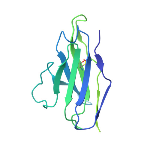
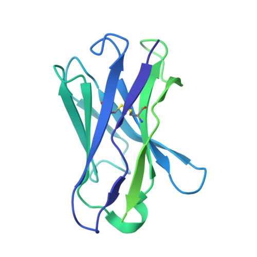
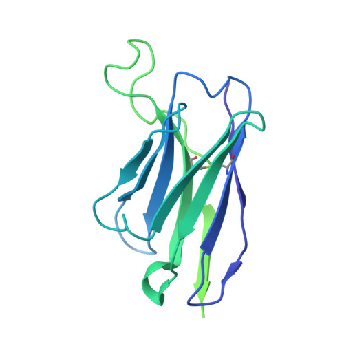
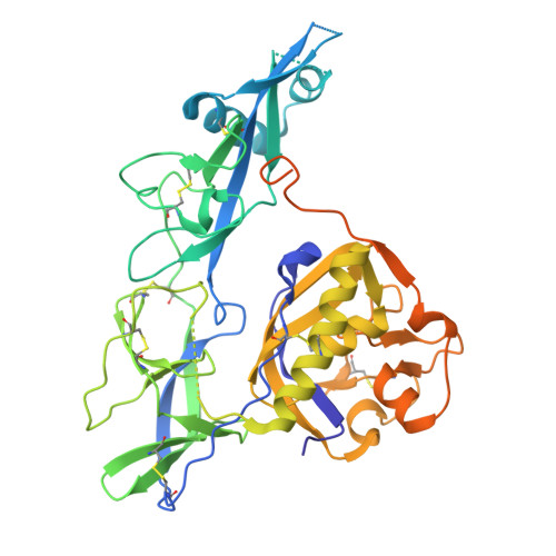
Rabies continues to kill over 60,000 people per year despite life-saving vaccines and post-exposure treatments and costs billions of dollars in prevention and treatment. Preventing rabies deaths and reducing the global economic burden of the virus will require both developing a monoclonal antibody cocktail to replace human serum in treatment and improving rabies vaccines to elicit long-lasting protection. Here, we solve nine cryo-electron microscopy (cryo-EM) structures of neutralizing monoclonal antibodies (mAbs) in complex with the rabies virus glycoprotein (RABV-G). The nine structures span three known antigenic sites plus two additional antigenic sites, not among the five classically identified sites. We find that these antigenic sites, V and VI, are broadly cross-reactive across lyssaviruses, whereas immunodominant sites II/IV and III are rabies specific. Across the mAb panel, fusion inhibition and binding affinity correlate best with neutralization. Together, these results provide a roadmap for structure-guided vaccine and therapeutic antibody design for rabies and related lyssaviruses.
- Center for Vaccine Innovation, La Jolla Institute for Immunology, La Jolla, CA 92037, USA; Department of Chemistry and Biochemistry, Montana State University, Bozeman, MT 59717, USA; Department of Microbiology and Cell Biology, Montana State University, Bozeman, MT 59717, USA. Electronic address: heather.callaway@montana.edu.
Organizational Affiliation: