 Download Mendeley
Cryo-EM structure of a methanogen nitrogenase-PII protein supercomplex
Kashyap, R., Deere, T.M., Dhamad, A., Chanderban, M., Tokmina-Lukaszewska, M., Bothner, B., Lessner, D.J., Antony, E.(2026) Nature
Experimental Data Snapshot
Starting Model: experimental
View more details
wwPDB Validation 3D Report Full Report

(2026) Nature
Entity ID: 1 | |||||
|---|---|---|---|---|---|
| Molecule | Chains | Sequence Length | Organism | Details | Image |
| Nitrogenase molybdenum-iron protein beta chain | A [auth B], B [auth D] | 456 | Methanosarcina acetivorans | Mutation(s): 0 EC: 1.18.6.1 | 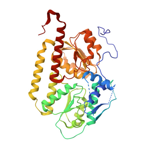 |
UniProt | |||||
Entity Groups | |||||
| Sequence Clusters | 30% Identity50% Identity70% Identity90% Identity95% Identity100% Identity | ||||
| UniProt Group | Q8TJ89 | ||||
Sequence AnnotationsExpand | |||||
Reference Sequence | |||||
Entity ID: 2 | |||||
|---|---|---|---|---|---|
| Molecule | Chains | Sequence Length | Organism | Details | Image |
| Nitrogenase protein alpha chain | C [auth A], D [auth C] | 559 | Methanosarcina acetivorans | Mutation(s): 0 EC: 1.18.6.1 | 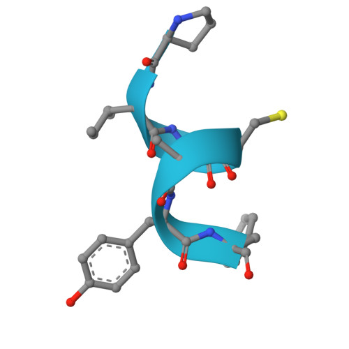 |
UniProt | |||||
Entity Groups | |||||
| Sequence Clusters | 30% Identity50% Identity70% Identity90% Identity95% Identity100% Identity | ||||
| UniProt Group | Q8TJ90 | ||||
Sequence AnnotationsExpand | |||||
Reference Sequence | |||||
| Ligands 1 Unique | |||||
|---|---|---|---|---|---|
| ID | Chains | Name / Formula / InChI Key | 2D Diagram | 3D Interactions | |
| CLF (Subject of Investigation/LOI) Download:Ideal Coordinates CCD File | E [auth B], F [auth D] | FE(8)-S(7) CLUSTER Fe8 S7 JKVMXLBGZBULKV-UHFFFAOYSA-N | 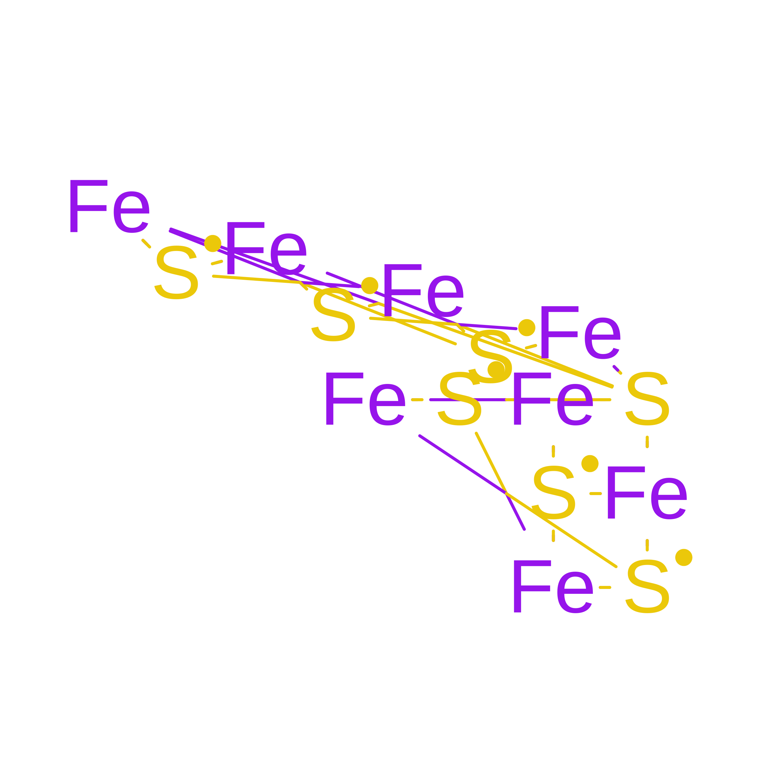 | ||
| Task | Software Package | Version |
|---|---|---|
| RECONSTRUCTION | PHENIX | 2.0-5936 |
| MODEL REFINEMENT | PHENIX | 2.0_5936 |
| Funding Organization | Location | Grant Number |
|---|---|---|
| Department of Energy (DOE, United States) | United States | DE-SC0020965 |
