11RJ | pdb_000011rj

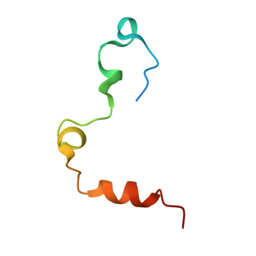
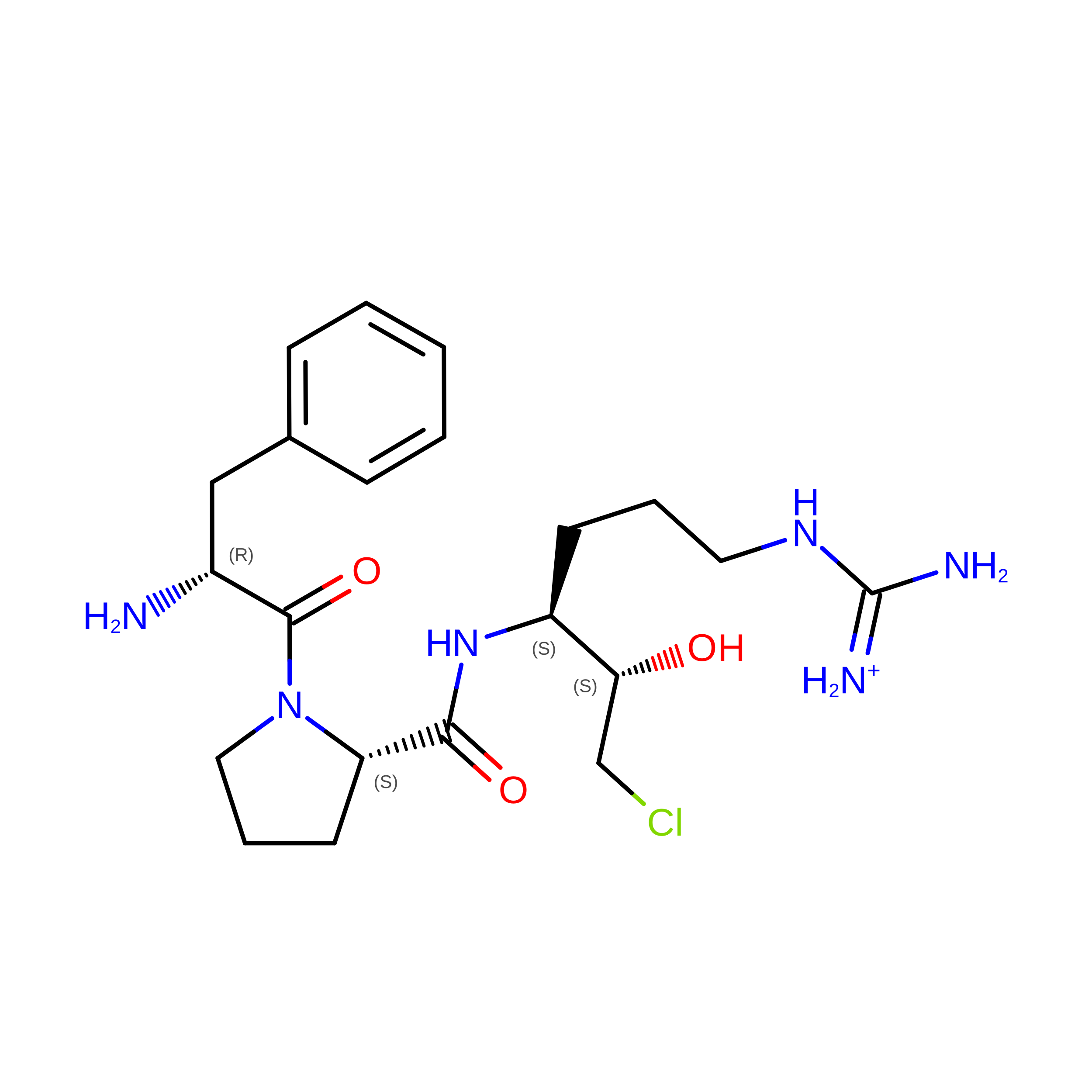
D189A thrombin inhibited with D-Phe-Pro-Arg-Chloromethylketone

  • Classification: BLOOD CLOTTING
  • Organism(s): Homo sapiens
  • Expression System: Mesocricetus auratus
  • Mutation(s): Yes 

  • Deposited: 2026-03-10 Released: 2026-10-07 
  • Deposition Author(s): Friet, T., Mohammed, B.M., Sukumar, N., Di Cera, E.
  • Funding Organization(s): National Institutes of Health/National Heart, Lung, and Blood Institute (NIH/NHLBI), National Institutes of Health/National Institute of General Medical Sciences (NIH/NIGMS), Department of Energy (DOE, United States)

Experimental Data Snapshot

  • Method: X-RAY DIFFRACTION
  • Resolution: 2.50 Å
  • R-Value Free: 
    0.287 (Depositor), 0.287 (DCC) 
  • R-Value Work: 
    0.229 (Depositor), 0.230 (DCC) 
  • R-Value Observed: 
    0.232 (Depositor) 

Starting Model: experimental
View more details

wwPDB Validation 3D Report Full Report

Validation slider image for 11RJ

Ligand Structure Quality Assessment 


This is version 1.0 of the entry. See complete history. 

Literature

Structural analysis of the primary specificity of thrombin.

Friet, T., Mikhail, G., Mohammed, B.M., Pelc, L.A., Dei Rossi, A., Korolev, S., Di Cera, E.

(2026) J Thromb Haemost 

  • DOI: https://doi.org/10.1016/j.jtha.2026.09.021
  • Primary Citation Related Structures: 
    11RI, 11RJ, 11RK, 11VG, 11XW, 11YH, 12CW, 12CX, 12CY, 12IT

  • PubMed Abstract: 

    Thrombin has dual trypsin-like and chymotrypsin-like specificity: it prefers substrates carrying Arg at the site of cleavage (P1) in the activation peptide because of the presence of D189 in the primary specificity (S1) site but can also cleave substrates carrying Phe at P1. Explore the structural basis of the P1-S1 interaction with mutants of D189 and substrates carrying different residues at P1. X-ray crystallography is used to solve the structures of thrombin wild-type and mutants D189A, D189F and D189K bound to the irreversible inhibitors H-D-Phe-Pro-Arg-CH 2 Cl (FPRck), H-D-Phe-Pro-Phe-CH 2 Cl (FPFck) and H-D-Phe-Pro-Gln-CH 2 Cl (FPQck). X-ray structures of thrombin wild-type and mutants D189A, D189F and D189K bound to FPRck, FPFck and FPQck are solved at high resolutions, from 1.2 Å to 2.5 Å. Mutations of D189 do not affect stability of free thrombin but shift the substrate preference from Arg to Phe at P1 and the stability of the inhibited complex from FPRck to FPFck. The structures reveal the flexibility of the S1 site in accommodating side chains of different chemical properties to optimize the P1-S1 interaction. In the D189K-FPRck complex, the inhibitor is trapped in an intermediate state covalently bound only to the catalytic H57 and with the Arg side chain positioned outside of the active site. The S1 site of thrombin features unexpected structural flexibility and can withstand the replacement of D189 with Ala, Phe or even Lys. The results set the stage for future studies aimed at re-engineering primary specificity in thrombin and other trypsin-like proteases.


  • Organizational Affiliation: 
    • Edward A. Doisy Department of Biochemistry and Molecular Biology, Saint Louis University School of Medicine, St. Louis, MO 63104 USA.

Macromolecule Content 

  • Total Structure Weight: 37.5 kDa 
  • Atom Count: 2,510 
  • Modeled Residue Count: 295 
  • Deposited Residue Count: 322 
  • Unique protein chains: 2

Macromolecules

Find similar proteins by:|  3D Structure
Entity ID: 1
MoleculeChains  Sequence LengthOrganismDetailsImage
Thrombin heavy chainA [auth B]273Homo sapiensMutation(s): 1 
Gene Names: F2
EC: 3.4.21.5
UniProt & NIH Common Fund Data Resources
Find proteins for P00734 (Homo sapiens)
Explore P00734 
Go to UniProtKB:  P00734
PHAROS:  P00734
GTEx:  ENSG00000180210 
Entity Groups
Sequence Clusters30% Identity50% Identity70% Identity90% Identity95% Identity100% Identity
UniProt GroupP00734
Sequence Annotations
Expand
Reference Sequence
Find similar proteins by:|  3D Structure
Entity ID: 2
MoleculeChains  Sequence LengthOrganismDetailsImage
Thrombin light chainB [auth A]49Homo sapiensMutation(s): 0 
Gene Names: F2
EC: 3.4.21.5
UniProt & NIH Common Fund Data Resources
Find proteins for P00734 (Homo sapiens)
Explore P00734 
Go to UniProtKB:  P00734
PHAROS:  P00734
GTEx:  ENSG00000180210 
Entity Groups
Sequence Clusters30% Identity50% Identity70% Identity90% Identity95% Identity100% Identity
UniProt GroupP00734
Sequence Annotations
Expand
Reference Sequence

Small Molecules

Ligands 2 Unique
IDChains Name / Formula / InChI Key2D Diagram3D Interactions
0G6
(Subject of Investigation/LOI)

Query on 0G6


Download:Ideal Coordinates CCD File
C [auth B]D-phenylalanyl-N-[(2S,3S)-6-{[amino(iminio)methyl]amino}-1-chloro-2-hydroxyhexan-3-yl]-L-prolinamide
C21 H34 Cl N6 O3
DVFLYEYCMMLBTQ-VSZNYVQBSA-O
NA

Query on NA


Download:Ideal Coordinates CCD File
D [auth B]SODIUM ION
Na
FKNQFGJONOIPTF-UHFFFAOYSA-N

Biologically Interesting Molecules (External Reference) 

1 Unique

Experimental Data & Validation

Experimental Data

  • Method: X-RAY DIFFRACTION
  • Resolution: 2.50 Å
  • R-Value Free:  0.287 (Depositor), 0.287 (DCC) 
  • R-Value Work:  0.229 (Depositor), 0.230 (DCC) 
  • R-Value Observed: 0.232 (Depositor) 
Space Group: C 1 2 1
Unit Cell:
Length ( Å )Angle ( ˚ )
a = 134.113α = 90
b = 76.633β = 102.243
c = 44.869γ = 90
Software Package:
Software NamePurpose
PHENIXrefinement
Aimlessdata scaling
XDSdata reduction
PHASERphasing
PDB_EXTRACTdata extraction

Structure Validation

View Full Validation Report


Ligand Structure Quality Assessment 


Entry History 

& Funding Information

Deposition Data


Funding OrganizationLocationGrant Number
National Institutes of Health/National Heart, Lung, and Blood Institute (NIH/NHLBI)United StatesHL049413, HL139554, HL147821
National Institutes of Health/National Institute of General Medical Sciences (NIH/NIGMS)United StatesP30GM133893
Department of Energy (DOE, United States)United StatesKP1607011

Revision History  (Full details and data files)

  • Version 1.0: 2026-10-07
    Type: Initial release