11MP | pdb_000011mp

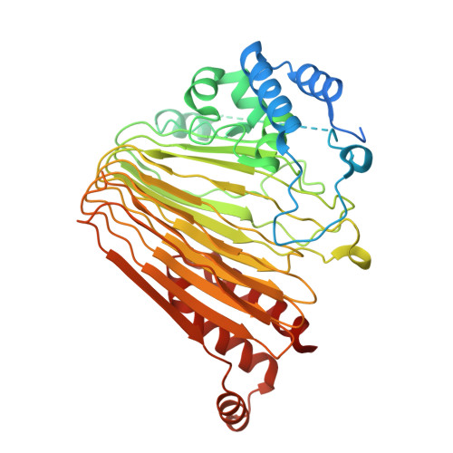
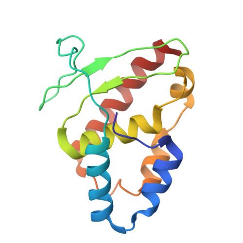
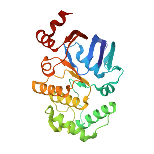
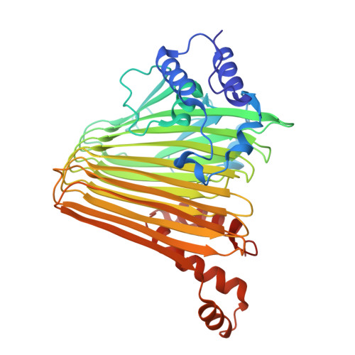
E. coli SufE bound to SufBC2D


Experimental Data Snapshot

  • Method: ELECTRON MICROSCOPY
  • Resolution: 4.21 Å
  • Aggregation State: PARTICLE 
  • Reconstruction Method: SINGLE PARTICLE 

wwPDB Validation 3D Report Full Report

Validation slider image for 11MP

This is version 1.0 of the entry. See complete history

Literature

The structure of the SufBC 2 D-SufE complex reveals the mechanism of sulfur transfer in bacterial Fe-S cluster assembly.

Chhikara, N.Mireku, N.Dunkle, J.A.Frantom, P.A.

(2026) bioRxiv 

  • DOI: https://doi.org/10.64898/2026.05.18.725997
  • Primary Citation Related Structures: 
    11MP

  • PubMed Abstract: 

    Iron-sulfur clusters are essential cofactors assembled in bacteria by the Suf pathway through a series of transient protein-protein interactions that transfer sulfur from L-cysteine to a scaffold complex. While early steps in persulfide transfer are well characterized, the mechanism of sulfur delivery to the SufBC 2 D scaffold has remained unresolved. Here, we report the first structure of the SufBC 2 D-SufE complex, capturing the final step in persulfide transfer in the Suf pathway. The structure reveals coordinated conformational changes in both SufB and SufE that expose the otherwise buried C254 acceptor site and position the SufE C51 loop beneath the SufB-SufD axis. Biochemical analysis of SufB variants demonstrates that substitutions in the globally conserved 220s β-strand enhance SufE binding affinity and persulfide transfer rates, consistent with stabilization of a locally rearranged, transfer-competent conformation. Together, these results support a model in which conformational gating regulates persulfide transfer, providing a mechanism for controlling access to reactive sulfur intermediates.


  • Organizational Affiliation
    • Department of Chemistry & Biochemistry, The University of Alabama, Tuscaloosa, AL, 35487, USA.

Macromolecule Content 

  • Total Structure Weight: 174.33 kDa 
  • Atom Count: 11,446 
  • Modeled Residue Count: 1,459 
  • Deposited Residue Count: 1,567 
  • Unique protein chains: 4

Macromolecules

Find similar proteins by:|  3D Structure
Entity ID: 1
MoleculeChains  Sequence LengthOrganismDetailsImage
Iron-sulfur cluster assembly protein SufBA [auth C]510Escherichia coliMutation(s): 1 
Gene Names: sufBynhEb1683JW5273
UniProt
Find proteins for P77522 (Escherichia coli (strain K12))
Explore P77522 
Go to UniProtKB:  P77522
Entity Groups
Sequence Clusters30% Identity50% Identity70% Identity90% Identity95% Identity100% Identity
UniProt GroupP77522
Sequence Annotations
Expand
Reference Sequence
Find similar proteins by:|  3D Structure
Entity ID: 2
MoleculeChains  Sequence LengthOrganismDetailsImage
Cysteine desulfuration protein SufEB [auth E]138Escherichia coliMutation(s): 0 
Gene Names: sufEynhAb1679JW1669
UniProt
Find proteins for P76194 (Escherichia coli (strain K12))
Explore P76194 
Go to UniProtKB:  P76194
Entity Groups
Sequence Clusters30% Identity50% Identity70% Identity90% Identity95% Identity100% Identity
UniProt GroupP76194
Sequence Annotations
Expand
Reference Sequence
Find similar proteins by:|  3D Structure
Entity ID: 3
MoleculeChains  Sequence LengthOrganismDetailsImage
Probable ATP-dependent transporter SufCC [auth A],
D [auth B]
248Escherichia coliMutation(s): 0 
Gene Names: sufCynhDb1682JW1672
UniProt
Find proteins for P77499 (Escherichia coli (strain K12))
Explore P77499 
Go to UniProtKB:  P77499
Entity Groups
Sequence Clusters30% Identity50% Identity70% Identity90% Identity95% Identity100% Identity
UniProt GroupP77499
Sequence Annotations
Expand
Reference Sequence
Find similar proteins by:|  3D Structure
Entity ID: 4
MoleculeChains  Sequence LengthOrganismDetailsImage
Iron-sulfur cluster assembly protein SufDE [auth D]423Escherichia coliMutation(s): 0 
Gene Names: sufDynhCb1681JW1671
UniProt
Find proteins for P77689 (Escherichia coli (strain K12))
Explore P77689 
Go to UniProtKB:  P77689
Entity Groups
Sequence Clusters30% Identity50% Identity70% Identity90% Identity95% Identity100% Identity
UniProt GroupP77689
Sequence Annotations
Expand
Reference Sequence

Experimental Data & Validation

Experimental Data

  • Method: ELECTRON MICROSCOPY
  • Resolution: 4.21 Å
  • Aggregation State: PARTICLE 
  • Reconstruction Method: SINGLE PARTICLE 
EM Software:
TaskSoftware PackageVersion
MODEL REFINEMENTPHENIX1.21.2_5419
RECONSTRUCTIONcryoSPARC4.6

Structure Validation

View Full Validation Report


Entry History 

& Funding Information

Deposition Data


Funding OrganizationLocationGrant Number
National Institutes of Health/National Institute of General Medical Sciences (NIH/NIGMS)United StatesR01GM112919
National Institutes of Health/National Institute of General Medical Sciences (NIH/NIGMS)United StatesR35GM142966

Revision History  (Full details and data files)

  • Version 1.0: 2026-07-15
    Type: Initial release