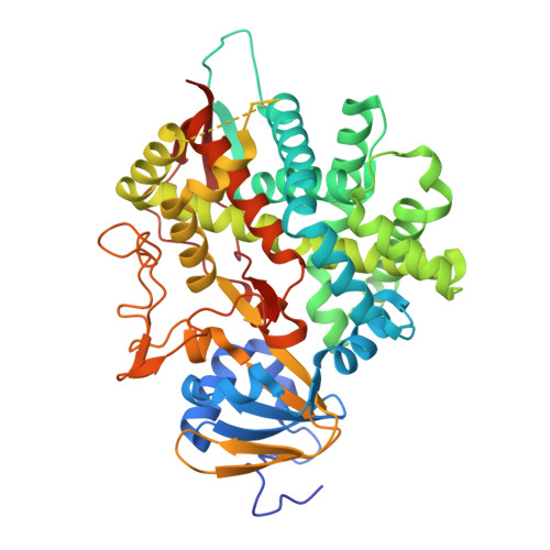
11IH | pdb_000011ih

CYP8B1 inhibitor compound 1


Experimental Data Snapshot

  • Method: X-RAY DIFFRACTION
  • Resolution: 1.70 Å
  • R-Value Free: 
    0.225 (Depositor), 0.222 (DCC) 
  • R-Value Work: 
    0.206 (Depositor), 0.202 (DCC) 
  • R-Value Observed: 
    0.206 (Depositor) 

Starting Model: experimental
View more details

wwPDB Validation 3D Report Full Report

Validation slider image for 11IH

Ligand Structure Quality Assessment 


This is version 1.0 of the entry. See complete history. 

Literature

Structure-Based Drug Design of Quinoline CYP8B1 Inhibitors for the Potential Treatment of Metabolic Disease.

McCracken, A.B., Li, D., Caldwell, J.P., Lankin, C., Tagat, J., Khan, T., Rao, A., Zhang, T., Caplen, M.A., Verras, A., Orth, P., Hruza, A., Ho, T., Allen, S., Elsen, N.L., Dingley, K., Duffy, R., Wisniewski, T., Murphy, B., Castro-Perez, J., Wong, K., Streckfuss, E., Liu, J., Tong, W., McKittrick, B.

(2026) ACS Med Chem Lett 17: 2245-2250

  • DOI: https://doi.org/10.1021/acsmedchemlett.6c00387
  • Primary Citation Related Structures: 
    11IH, 11IR

  • PubMed Abstract: 

    CYP8B1 has been identified as a potential target for the treatment of metabolic diseases, such as nonalcoholic fatty liver disease (NAFLD) and type 2 diabetes (T2D). CYP8B1 enzyme inhibitor 1 was discovered from a high-throughput screen of our compound collection; however, its poor metabolic stability prevented further in vivo evaluation. Leveraging structure-based drug design, metabolite identification and metabolic stability-guided DMPK optimization, and library synthesis to rapidly expand SAR, we identified compound 9 with substantially improved DMPK properties. Compound 9 showed robust in vivo CYP8B1 inhibition in mice and provided the foundation for further optimization of this class of CYP8B1 inhibitors.


  • Organizational Affiliation: 
    • Merck & Co., Inc., Rahway, New Jersey 07065, United States.

Macromolecule Content 

  • Total Structure Weight: 57.98 kDa 
  • Atom Count: 4,091 
  • Modeled Residue Count: 461 
  • Deposited Residue Count: 488 
  • Unique protein chains: 1

Macromolecules

Find similar proteins by:|  3D Structure
Entity ID: 1
MoleculeChains  Sequence LengthOrganismDetailsImage
7-alpha-hydroxycholest-4-en-3-one 12-alpha-hydroxylase488Homo sapiensMutation(s): 0 
Gene Names: CYP8B1, CYP12
EC: 1.14.14.139
UniProt & NIH Common Fund Data Resources
Find proteins for Q9UNU6 (Homo sapiens)
Explore Q9UNU6 
Go to UniProtKB:  Q9UNU6
PHAROS:  Q9UNU6
GTEx:  ENSG00000180432 
Entity Groups
Sequence Clusters30% Identity50% Identity70% Identity90% Identity95% Identity100% Identity
UniProt GroupQ9UNU6
Sequence Annotations
Expand
Reference Sequence

Small Molecules

Ligands 2 Unique
IDChains Name / Formula / InChI Key2D Diagram3D Interactions
HEM

Query on HEM


Download:Ideal Coordinates CCD File
B [auth A]PROTOPORPHYRIN IX CONTAINING FE
C34 H32 Fe N4 O4
KABFMIBPWCXCRK-RGGAHWMASA-L
A1C9D(
Subject of Investigation/LOI)

Query on A1C9D


Download:Ideal Coordinates CCD File
C [auth A]6-methyl-4-[4-(4-methylpiperidine-1-carbonyl)piperidin-1-yl]quinoline-3-carbonitrile
C23 H28 N4 O
BJGUYZVGGPNEGT-UHFFFAOYSA-N

Experimental Data & Validation

Experimental Data

  • Method: X-RAY DIFFRACTION
  • Resolution: 1.70 Å
  • R-Value Free:  0.225 (Depositor), 0.222 (DCC) 
  • R-Value Work:  0.206 (Depositor), 0.202 (DCC) 
  • R-Value Observed: 0.206 (Depositor) 
Space Group: P 21 21 21
Unit Cell:
Length ( Å )Angle ( ˚ )
a = 57.844α = 90
b = 89.708β = 90
c = 106.436γ = 90
Software Package:
Software NamePurpose
BUSTERrefinement
DENZOdata reduction
SCALEPACKdata scaling
REFMACphasing

Structure Validation

View Full Validation Report


Ligand Structure Quality Assessment 


Entry History 

& Funding Information

Deposition Data


Funding OrganizationLocationGrant Number
Not funded--

Revision History  (Full details and data files)

  • Version 1.0: 2026-10-28
    Type: Initial release