11AD | pdb_000011ad

Structural basis for high-affinity inhibitor binding to lipid kinases PIP4K2A and PIP4K2B

  • Classification: TRANSFERASE
  • Organism(s): Homo sapiens
  • Expression System: Escherichia coli
  • Mutation(s): No 

  • Deposited: 2026-02-13 Released: 2026-04-29 
  • Deposition Author(s): He, Z., Ha, Y.
  • Funding Organization(s): National Institutes of Health/National Institute of General Medical Sciences (NIH/NIGMS)

Experimental Data Snapshot

  • Method: X-RAY DIFFRACTION
  • Resolution: 2.79 Å
  • R-Value Free: 
    0.234 (Depositor), 0.233 (DCC) 
  • R-Value Work: 
    0.169 (Depositor), 0.169 (DCC) 
  • R-Value Observed: 
    0.173 (Depositor) 

Starting Model: experimental
View more details

wwPDB Validation 3D Report Full Report

Validation slider image for 11AD

Ligand Structure Quality Assessment 


This is version 1.1 of the entry. See complete history

Literature

Structural basis for high-affinity inhibitor binding to lipid kinases PIP4K2A and PIP4K2B.

He, Z.Chen, S.Micheli, F.Cianciulli, A.Beato, C.Ellman, J.Ha, Y.

(2026) Acta Crystallogr D Struct Biol 82: 646-654

  • DOI: https://doi.org/10.1107/S2059798326003773
  • Primary Citation Related Structures: 
    11AD

  • PubMed Abstract: 

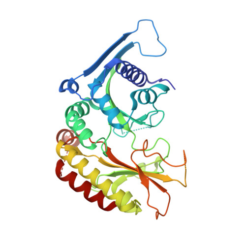
    The phosphatidylinositol 5-phosphate 4-kinases (PIP4Ks) are an evolutionarily conserved family of lipid kinases that phosphorylate phosphatidylinositol 5-phosphate to generate phosphatidylinositol 4,5-bisphosphate. In mammals, the catalytically active α and β isoforms, encoded by PIP4K2A and PIP4K2B, respectively, localize to distinct cellular compartments and have been implicated in metabolism, immune regulation and tumorigenesis, prompting interest in their pharmacological inhibition. Notably, most reported small-molecule inhibitors display substantially higher potency towards the α isoform than the β isoform, suggesting intrinsic structural features that limit the effective targeting of PIP4K2B. Here, we report the crystal structure of PIP4K2A in complex with 422A, a potent dual α/β inhibitor with improved metabolic stability. The structure reveals an unexpected, water-mediated interaction in which a pyridyl nitrogen of the inhibitor engages a conserved structured water molecule in the roof of the specificity pocket, constraining the orientation of the pyridylmethyl side chain and stabilizing a rigid, high-affinity binding mode. Comparative structural analysis with the PIP4K2A-selective inhibitor BAY-091 shows that deeper penetration into the specificity pocket enhances PIP4K2A binding but is accompanied by local steric constraints that are likely to be less well tolerated in PIP4K2B. Together, these findings define structural determinants of isoform-dependent inhibitor binding within the PIP4K family and provide a framework for structure-guided optimization of lipid kinase inhibitors with improved isoform balance.


  • Organizational Affiliation
    • Department of Pharmacology, Yale School of Medicine, New Haven, CT 06520, USA.

Macromolecule Content 

  • Total Structure Weight: 43.9 kDa 
  • Atom Count: 2,751 
  • Modeled Residue Count: 317 
  • Deposited Residue Count: 377 
  • Unique protein chains: 1

Macromolecules

Find similar proteins by:|  3D Structure
Entity ID: 1
MoleculeChains  Sequence LengthOrganismDetailsImage
Phosphatidylinositol 5-phosphate 4-kinase type-2 alpha377Homo sapiensMutation(s): 0 
Gene Names: PIP4K2API5P4KAPIP5K2PIP5K2A
EC: 2.7.1.149
UniProt & NIH Common Fund Data Resources
Find proteins for P48426 (Homo sapiens)
Explore P48426 
Go to UniProtKB:  P48426
PHAROS:  P48426
GTEx:  ENSG00000150867 
Entity Groups
Sequence Clusters30% Identity50% Identity70% Identity90% Identity95% Identity100% Identity
UniProt GroupP48426
Sequence Annotations
Expand
Reference Sequence

Experimental Data & Validation

Experimental Data

  • Method: X-RAY DIFFRACTION
  • Resolution: 2.79 Å
  • R-Value Free:  0.234 (Depositor), 0.233 (DCC) 
  • R-Value Work:  0.169 (Depositor), 0.169 (DCC) 
  • R-Value Observed: 0.173 (Depositor) 
Space Group: P 61 2 2
Unit Cell:
Length ( Å )Angle ( ˚ )
a = 135.883α = 90
b = 135.883β = 90
c = 94.679γ = 120
Software Package:
Software NamePurpose
REFMACrefinement
PDB_EXTRACTdata extraction
HKL-2000data reduction
HKL-2000data scaling
REFMACphasing

Structure Validation

View Full Validation Report


Ligand Structure Quality Assessment 


Entry History 

& Funding Information

Deposition Data

  • Released Date: 2026-04-29 
  • Deposition Author(s): He, Z., Ha, Y.

Funding OrganizationLocationGrant Number
National Institutes of Health/National Institute of General Medical Sciences (NIH/NIGMS)United StatesGM138722
National Institutes of Health/National Institute of General Medical Sciences (NIH/NIGMS)United StatesGM150502

Revision History  (Full details and data files)

  • Version 1.0: 2026-04-29
    Type: Initial release
  • Version 1.1: 2026-06-10
    Changes: Database references