10GI | pdb_000010gi

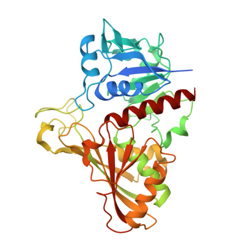
Structure of Glyceraldehyde-3-Phosphate Dehydrogenase from Babesia bovis


Experimental Data Snapshot

  • Method: X-RAY DIFFRACTION
  • Resolution: 3.12 Å
  • R-Value Free: 
    0.341 (Depositor), 0.340 (DCC) 
  • R-Value Work: 
    0.290 (Depositor), 0.290 (DCC) 
  • R-Value Observed: 
    0.293 (Depositor) 

Starting Model: in silico
View more details

wwPDB Validation 3D Report Full Report

Validation slider image for 10GI

Ligand Structure Quality Assessment 


This is version 1.1 of the entry. See complete history

Literature

Expression, purification, crystallization and structure solution of glyceraldehyde-3-phosphate dehydrogenase from the babesiosis infective agent Babesia bovis.

Ukrainski, B.Galvao, E.B.Silva, M.Iulek, J.

(2026) Protein Expr Purif 243: 106991-106991

  • DOI: https://doi.org/10.1016/j.pep.2026.106991
  • Primary Citation Related Structures: 
    10GI

  • PubMed Abstract: 

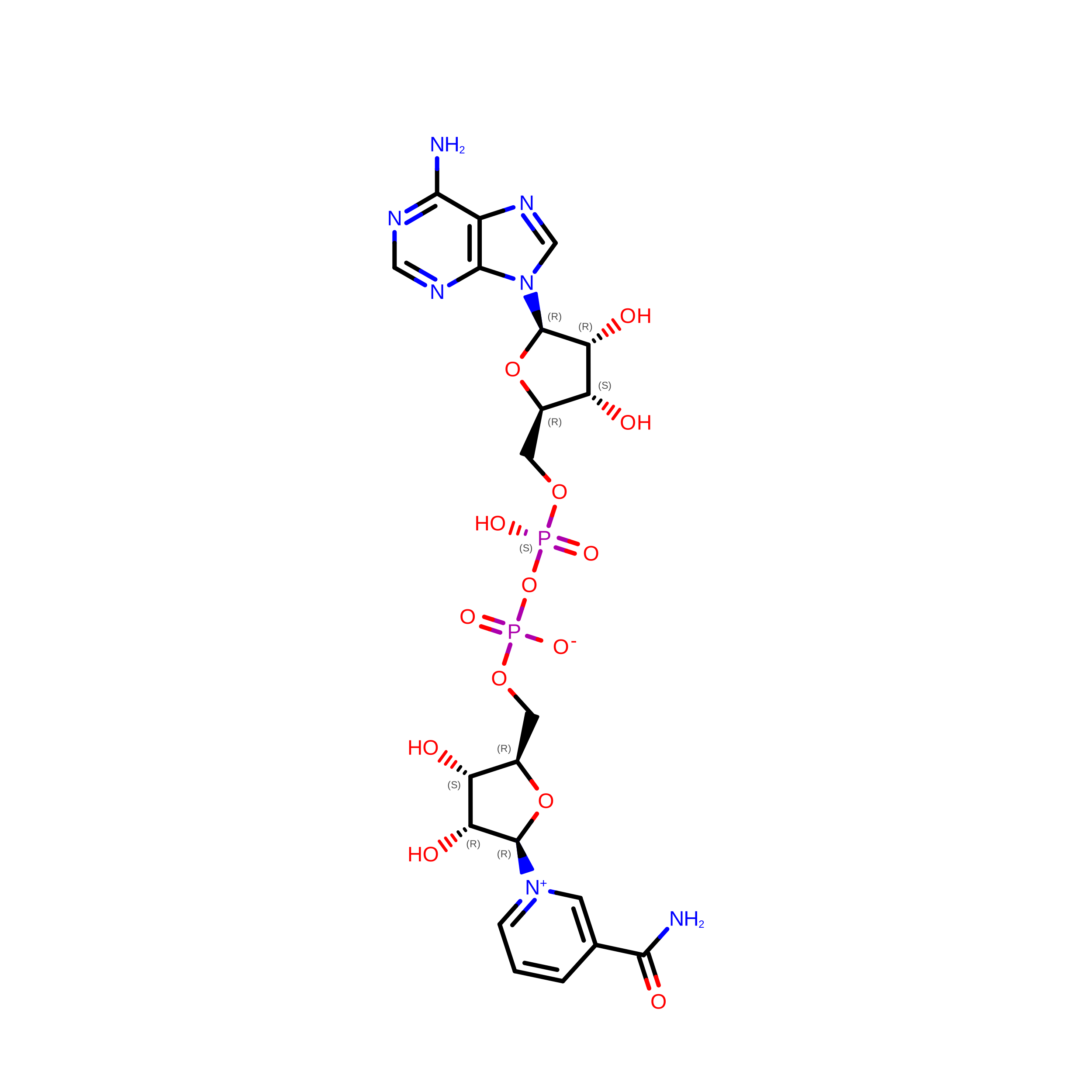
    Babesia bovis is a species of apicomplexan hemoparasitic protozoa that can be transmitted by ticks, causing a global cattle disease. As it depends mainly on the glycolytic pathway for energy production and life cycle maintenance, glycolytic enzymes are possible targets for drug development against Babesia. Glyceraldehyde-3-Phosphate Dehydrogenase (GAPDH) has been one of such targets, against several parasitic organisms. It performs the reversible oxidative phosphorylation of glyceraldehyde-3-phosphate to 1,3-bisphospho-D-glycerate in the presence of nicotinamide adenine dinucleotide. The protocol for Babesia bovis Glyceraldehyde-3-Phosphate Dehydrogenase (BbGAPDH) expression and purification has been developed to yields of 28 mg of pure protein per liter of culture medium, with a specific activity of 55.5 ± 6.99 U mg -1 after His-tag removal, indicating preservation of enzymatic activity. For crystallization, the His-tag removal proved essential. Crystals diffracted to 3.12 Å resolution in the P3 1 21 space group; NAD + cofactor molecules are observed in their respective sites. Comparisons to the theoretical model indicate a number of side chain conformation differences. These results provide support for future enzyme inhibition assays, in addition to crystallization assays with potential inhibitors.


  • Organizational Affiliation
    • Chemistry Departament, State University of Ponta Grossa, Paraná, Brazil. Electronic address: ukrainskibruna3@gmail.com.

Macromolecule Content 

  • Total Structure Weight: 78.33 kDa 
  • Atom Count: 5,074 
  • Modeled Residue Count: 675 
  • Deposited Residue Count: 716 
  • Unique protein chains: 1

Macromolecules

Find similar proteins by:|  3D Structure
Entity ID: 1
MoleculeChains  Sequence LengthOrganismDetailsImage
Glyceraldehyde-3-phosphate dehydrogenase
A, B
358Babesia bovisMutation(s): 0 
Gene Names: BBOV_II002540
EC: 1.2.1.12
UniProt
Find proteins for A7ATE8 (Babesia bovis)
Explore A7ATE8 
Go to UniProtKB:  A7ATE8
Entity Groups
Sequence Clusters30% Identity50% Identity70% Identity90% Identity95% Identity100% Identity
UniProt GroupA7ATE8
Sequence Annotations
Expand
Reference Sequence

Experimental Data & Validation

Experimental Data

  • Method: X-RAY DIFFRACTION
  • Resolution: 3.12 Å
  • R-Value Free:  0.341 (Depositor), 0.340 (DCC) 
  • R-Value Work:  0.290 (Depositor), 0.290 (DCC) 
  • R-Value Observed: 0.293 (Depositor) 
Space Group: P 31 2 1
Unit Cell:
Length ( Å )Angle ( ˚ )
a = 85.614α = 90
b = 85.614β = 90
c = 155.063γ = 120
Software Package:
Software NamePurpose
PHENIXrefinement
DIALSdata reduction
Aimlessdata scaling
PHASERphasing

Structure Validation

View Full Validation Report


Ligand Structure Quality Assessment 


Entry History 

& Funding Information

Deposition Data


Funding OrganizationLocationGrant Number
Other governmentBrazilPBA2025201000175

Revision History  (Full details and data files)

  • Version 1.0: 2026-08-19
    Type: Initial release
  • Version 1.1: 2026-08-26
    Changes: Database references