10DJ | pdb_000010dj

Fyn Kinase Domain-Saracatinib Complex Structure


Experimental Data Snapshot

  • Method: X-RAY DIFFRACTION
  • Resolution: 2.22 Å
  • R-Value Free: 
    0.268 (Depositor), 0.270 (DCC) 
  • R-Value Work: 
    0.245 (Depositor), 0.246 (DCC) 
  • R-Value Observed: 
    0.246 (Depositor) 

Starting Model: experimental
View more details

wwPDB Validation 3D Report Full Report

Validation slider image for 10DJ

Ligand Structure Quality Assessment 


This is version 1.1 of the entry. See complete history

Literature

Fyn-Saracatinib Complex Structure Reveals an Active State-like Conformation.

Ta, H.M.Sankaran, B.Roush, E.D.Ferreon, J.C.Ferreon, A.C.M.Kim, C.

(2026) Int J Mol Sci 27

  • DOI: https://doi.org/10.3390/ijms27031143
  • Primary Citation Related Structures: 
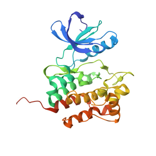
    10DJ

  • PubMed Abstract: 

    Fyn is a Src-family tyrosine kinase implicated in synaptic dysfunction and neuroinflammation across multiple neurodegenerative disorders, including Alzheimer's disease (AD) and Parkinson's disease (PD). Saracatinib (AZD0530) is a potent Src-family inhibitor that has been explored as a repurposed therapeutic; however, its clinical utility is limited by poor kinase selectivity caused by high sequence conservation within Src-family ATP-binding sites. Here, we combine surface plasmon resonance (SPR) and X-ray crystallography to define saracatinib recognition by the Fyn kinase domain (KD). SPR single-cycle kinetics shows that saracatinib binds the isolated Fyn KD and full-length Fyn with low-nanomolar affinity, whereas dasatinib binds with subnanomolar affinity and markedly slower dissociation. We determined the crystal structure of the Fyn KD-saracatinib complex at 2.22 Å resolution. The kinase adopts an active-like conformation with the DFG motif and αC-helix in the 'in' state and a conserved β3 αC Lys-Glu salt bridge. Saracatinib occupies the adenine and ribose pockets, and engages the hinge through direct and water-mediated hydrogen bonding while complementing a hydrophobic back pocket by van der Waals contacts. Comparison with reported saracatinib-bound structures of other kinases suggests that the active-state geometry observed for Fyn creates a pocket not observed in inactive-like complexes, providing a structural handle for designing Fyn-selective inhibitors. Comparison with all saracatinib-bound kinase co-structures currently available in the PDB (ALK2 and PKMYT1) indicates a conserved monodentate hinge binding mode but kinase-dependent αC-helix conformations, providing a structural rationale for designing Fyn-selective analogues.


  • Organizational Affiliation
    • Verna and Marrs McLean Department of Biochemistry and Molecular Pharmacology, Baylor College of Medicine, Houston, TX 77030, USA.

Macromolecule Content 

  • Total Structure Weight: 65.83 kDa 
  • Atom Count: 4,866 
  • Modeled Residue Count: 546 
  • Deposited Residue Count: 564 
  • Unique protein chains: 1

Macromolecules

Find similar proteins by:|  3D Structure
Entity ID: 1
MoleculeChains  Sequence LengthOrganismDetailsImage
Tyrosine-protein kinase Fyn
A, B
282Homo sapiensMutation(s): 0 
Gene Names: FYN
EC: 2.7.10.2
UniProt & NIH Common Fund Data Resources
Find proteins for P06241 (Homo sapiens)
Explore P06241 
Go to UniProtKB:  P06241
PHAROS:  P06241
GTEx:  ENSG00000010810 
Entity Groups
Sequence Clusters30% Identity50% Identity70% Identity90% Identity95% Identity100% Identity
UniProt GroupP06241
Sequence Annotations
Expand
Reference Sequence

Small Molecules

Ligands 1 Unique
IDChains Name / Formula / InChI Key2D Diagram3D Interactions
H8H
(Subject of Investigation/LOI)

Query on H8H


Download:Ideal Coordinates CCD File
C [auth A],
D [auth B]
N-(5-CHLORO-1,3-BENZODIOXOL-4-YL)-7-[2-(4-METHYLPIPERAZIN-1-YL)ETHOXY]-5-(TETRAHYDRO-2H-PYRAN-4-YLOXY)QUINAZOLIN-4-AMINE
C27 H32 Cl N5 O5
OUKYUETWWIPKQR-UHFFFAOYSA-N

Experimental Data & Validation

Experimental Data

  • Method: X-RAY DIFFRACTION
  • Resolution: 2.22 Å
  • R-Value Free:  0.268 (Depositor), 0.270 (DCC) 
  • R-Value Work:  0.245 (Depositor), 0.246 (DCC) 
  • R-Value Observed: 0.246 (Depositor) 
Space Group: P 21 21 2
Unit Cell:
Length ( Å )Angle ( ˚ )
a = 79.687α = 90
b = 89.601β = 90
c = 92.01γ = 90
Software Package:
Software NamePurpose
PHENIXrefinement
XSCALEdata scaling
HKL-2000data reduction
PHENIXphasing
PDB_EXTRACTdata extraction

Structure Validation

View Full Validation Report


Ligand Structure Quality Assessment 


Entry History 

& Funding Information

Deposition Data


Funding OrganizationLocationGrant Number
National Institutes of Health/National Institute of General Medical Sciences (NIH/NIGMS)United States--

Revision History  (Full details and data files)

  • Version 1.0: 2026-03-18
    Type: Initial release
  • Version 1.1: 2026-03-25
    Changes: Database references