Primary Citation of Related Structures: 7ZJW, 7ZJX
PubMed Abstract:
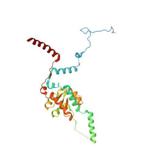
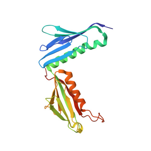
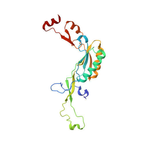
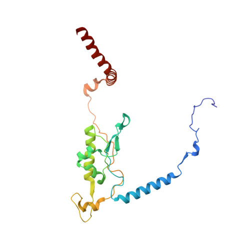
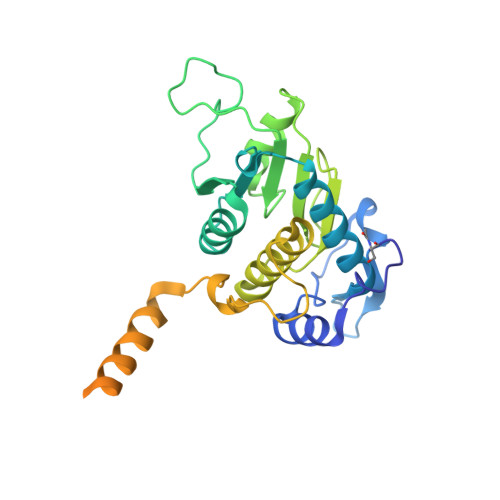
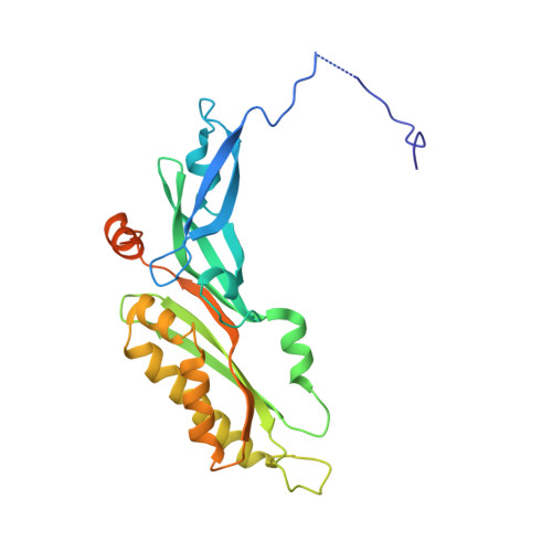
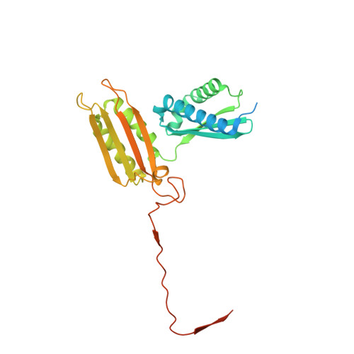
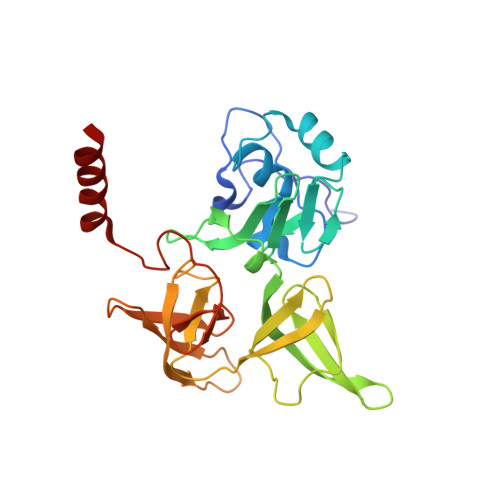
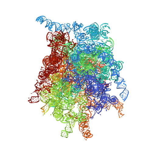
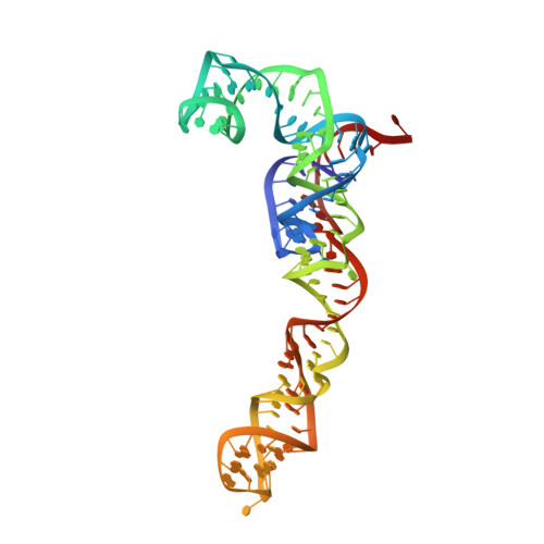
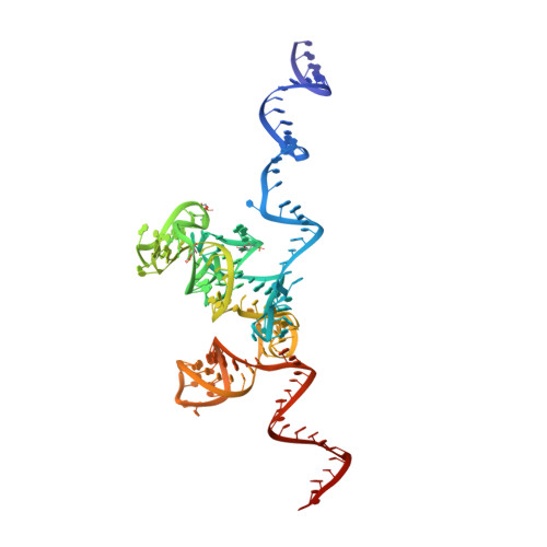
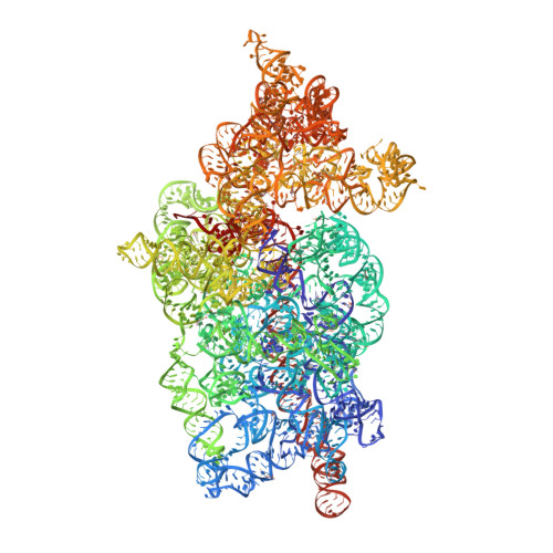
The elongation of eukaryotic selenoproteins relies on a poorly understood process of interpreting in-frame UGA stop codons as selenocysteine (Sec). We used cryo-electron microscopy to visualize Sec UGA recoding in mammals. A complex between the noncoding Sec-insertion sequence (SECIS), SECIS-binding protein 2 (SBP2), and 40 S ribosomal subunit enables Sec-specific elongation factor eEFSec to deliver Sec. eEFSec and SBP2 do not interact directly but rather deploy their carboxyl-terminal domains to engage with the opposite ends of the SECIS. By using its Lys-rich and carboxyl-terminal segments, the ribosomal protein eS31 simultaneously interacts with Sec-specific transfer RNA (tRNA Sec ) and SBP2, which further stabilizes the assembly. eEFSec is indiscriminate toward l-serine and facilitates its misincorporation at Sec UGA codons. Our results support a fundamentally distinct mechanism of Sec UGA recoding in eukaryotes from that in bacteria.
Organizational Affiliation:
Institut für Medizinische Physik und Biophysik, Charité-Universitätsmedizin Berlin, 10117 Berlin, Germany.
AJ [auth L5] BJ [auth L5] CJ [auth L5] DJ [auth L5] EJ [auth L5]
AJ [auth L5], BJ [auth L5], CJ [auth L5], DJ [auth L5], EJ [auth L5], FJ [auth L5], GJ [auth L5], HJ [auth L5], IJ [auth L5], SI [auth L5], TI [auth L5], UI [auth L5], VI [auth L5], WI [auth L5], XI [auth L5], YI [auth L5], YL [auth S2], ZI [auth L5]