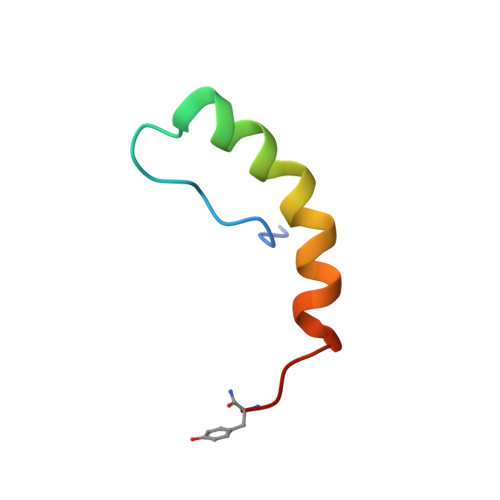
Structural similarities of micelle-bound peptide YY (PYY) and neuropeptide Y (NPY) are related to their affinity profiles at the Y receptors.
Lerch, M., Mayrhofer, M., Zerbe, O.(2004) J Mol Biol 339: 1153-1168
- PubMed: 15178255
- DOI: https://doi.org/10.1016/j.jmb.2004.04.032
- Primary Citation of Related Structures:
1RU5, 1RUU - PubMed Abstract:
Here, we investigate the structure of porcine peptide YY (pPYY) both when unligated in solution at pH 4.2 and when bound to dodecylphosphocholine (DPC) micelles at pH 5.5. pPYY in solution displays the PP-fold, with the N-terminal segment being back-folded onto the C-terminal alpha-helix, which extends from residue 17 to 31. In contrast to the solution structure of Keire et al. published in the year 2000 the C-terminal helix does not display a kink around residue 23-25. The root mean square deviation (RMSD) for backbone atoms of the NMR ensemble of conformers to the mean structure is 0.99(+/-0.35) Angstrom for residues 14-31. The back-fold is supported by values of 0.60+/-0.1 for the (15)N(1)H-NOE and by generalized order parameters S(2) of 0.74+/-0.1 for residues 5-31 which indicate that the peptide is folded in that segment. We have additionally used DPC micelles as a membrane model and determined the structure of pPYY when bound to it. Therein, an alpha-helix occurs in the segment comprising residues 17-31 and the N terminus freely diffuses in solution. The hydrophobic side of the amphipathic helix forms the micelle-binding interface and hydrophobic side-chains extend into the micelle interior. A significant stabilization of helical conformation occurs in the C-terminal pentapeptide, which is important for receptor binding. The latter is supported by positive values of the heteronuclear NOE in that segment (0.52+/-0.1 compared to 0.08+/-0.4 for the unligated form) and by values of S(2) of 0.6+/-0.2 (versus 0.38+/-0.2 for the unligated form). The structures of micelle-bound pPYY and pNPY are much more similar than those of pPYY and bPP with pairwise RMSDs of 1.23(+/-0.21)A or 3.21(+/-0.39) Angstrom, respectively. In contrast to the conformational similarities in the DPC-bound state their structures in solution are very different. In fact pPYY is more similar to bPP, which with its strong preference for the Y(4) receptor displays a completely different binding profile. Considering the high degree of sequence homology of pNPY and pPYY (>80%) and the fact, that their binding affinities at all receptor subtypes are high and, more importantly, rather similar, it is much more likely that PYY and NPY are recognized by the Y receptors from the membrane-bound state. As a consequence of the latter the PP-fold is not important for recognition of PYY or NPY at the Y receptors. To our knowledge this work provides for the first time strong arguments derived from structural data that support a membrane-bound receptor recognition pathway.
Organizational Affiliation:
Institute of Pharmaceutical Sciences, ETH Zurich, Winterthurerstrasse 190, CH 8057 Zurich, Switzerland.


