


Crystal structure of CotB2 variant V80L
Dimos, N., Himpich, S., Driller, R., Loll, B.To be published.
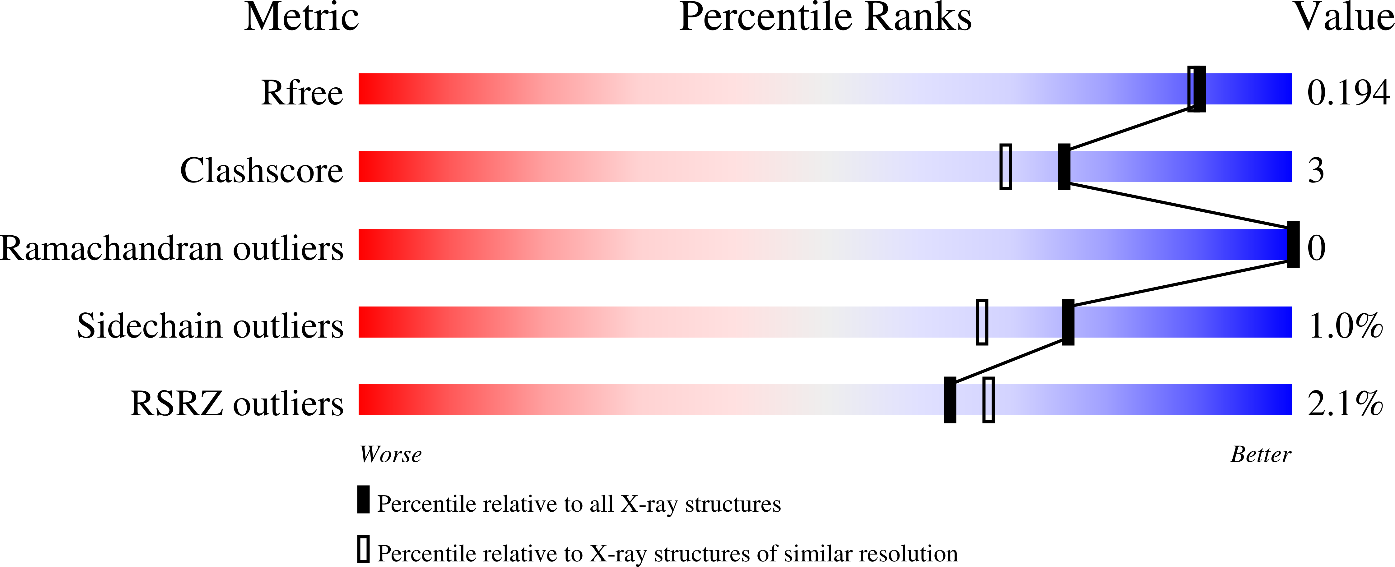
Experimental Data Snapshot
Entity ID: 1 | |||||
|---|---|---|---|---|---|
| Molecule | Chains | Sequence Length | Organism | Details | Image |
| Cyclooctat-9-en-7-ol synthase | 318 | Streptomyces melanosporofaciens | Mutation(s): 1 Gene Names: CotB2 | 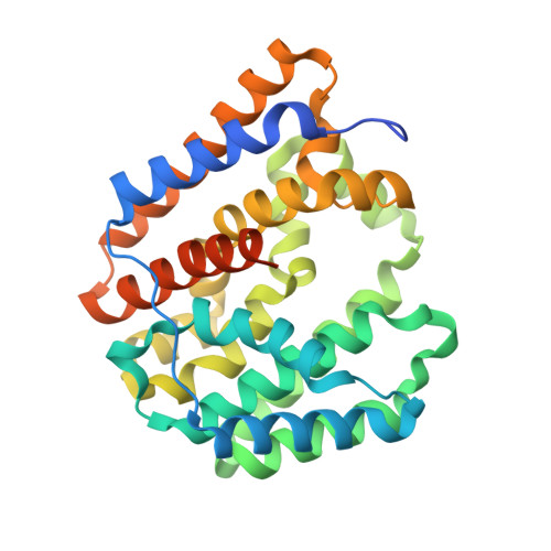 | |
UniProt | |||||
Find proteins for C9K1X5 (Streptomyces melanosporofaciens) Explore C9K1X5 Go to UniProtKB: C9K1X5 | |||||
Entity Groups | |||||
| Sequence Clusters | 30% Identity50% Identity70% Identity90% Identity95% Identity100% Identity | ||||
| UniProt Group | C9K1X5 | ||||
Sequence AnnotationsExpand | |||||
| |||||
| Ligands 3 Unique | |||||
|---|---|---|---|---|---|
| ID | Chains | Name / Formula / InChI Key | 2D Diagram | 3D Interactions | |
| CXS Query on CXS | C [auth A], F [auth B] | 3-CYCLOHEXYL-1-PROPYLSULFONIC ACID C9 H19 N O3 S PJWWRFATQTVXHA-UHFFFAOYSA-N |  | ||
| MPD Query on MPD | E [auth A], H [auth B] | (4S)-2-METHYL-2,4-PENTANEDIOL C6 H14 O2 SVTBMSDMJJWYQN-YFKPBYRVSA-N |  | ||
| NA Query on NA | D [auth A], G [auth B] | SODIUM ION Na FKNQFGJONOIPTF-UHFFFAOYSA-N |  | ||
| Length ( Å ) | Angle ( ˚ ) |
|---|---|
| a = 59.32 | α = 90 |
| b = 100.57 | β = 90 |
| c = 108.75 | γ = 90 |
| Software Name | Purpose |
|---|---|
| PHENIX | refinement |
| XDS | data reduction |
| XSCALE | data scaling |
| PHASER | phasing |
| Funding Organization | Location | Grant Number |
|---|---|---|
| German-Israeli Foundation for Research and Development | Germany | I-85-302.5-2019 |
RCSB PDB (citation) is hosted by
RCSB PDB is a member of the

