


Structure of YtoQ
Garbers, T.B., Neumann, P., Ficner, R.To be published.
Experimental Data Snapshot
wwPDB Validation 3D Report Full Report

Entity ID: 1 | |||||
|---|---|---|---|---|---|
| Molecule | Chains | Sequence Length | Organism | Details | Image |
| YtoQ | 170 | Bacillus subtilis subsp. subtilis str. 168 | Mutation(s): 0 Gene Names: ytoQ, BSU29850 | 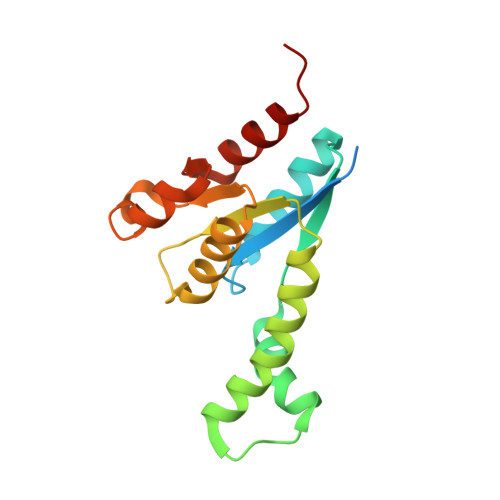 | |
UniProt | |||||
Find proteins for O34305 (Bacillus subtilis (strain 168)) Explore O34305 Go to UniProtKB: O34305 | |||||
Entity Groups | |||||
| Sequence Clusters | 30% Identity50% Identity70% Identity90% Identity95% Identity100% Identity | ||||
| UniProt Group | O34305 | ||||
Sequence AnnotationsExpand | |||||
| |||||
| Ligands 1 Unique | |||||
|---|---|---|---|---|---|
| ID | Chains | Name / Formula / InChI Key | 2D Diagram | 3D Interactions | |
| SO4 Query on SO4 | Q [auth B], R [auth C] | SULFATE ION O4 S QAOWNCQODCNURD-UHFFFAOYSA-L |  | ||
| Length ( Å ) | Angle ( ˚ ) |
|---|---|
| a = 137.34 | α = 90 |
| b = 70.15 | β = 98.91 |
| c = 149.94 | γ = 90 |
| Software Name | Purpose |
|---|---|
| PHENIX | refinement |
| XSCALE | data scaling |
| XDS | data reduction |
| PHASER | phasing |
| Funding Organization | Location | Grant Number |
|---|---|---|
| German Research Foundation (DFG) | Germany | -- |
RCSB PDB (citation) is hosted by
RCSB PDB is a member of the

