8K4R

Structure of VinM-VinL complex


Experimental Data Snapshot

  • Method: X-RAY DIFFRACTION
  • Resolution: 2.30 Å
  • R-Value Free: 0.251 
  • R-Value Work: 0.207 
  • R-Value Observed: 0.209 

wwPDB Validation   3D Report Full Report


Ligand Structure Quality Assessment 


This is version 1.1 of the entry. See complete history


Literature

Structural Basis of Amide-Forming Adenylation Enzyme VinM in Vicenistatin Biosynthesis.

Miyanaga, A.Nagata, K.Nakajima, J.Chisuga, T.Kudo, F.Eguchi, T.

(2023) ACS Chem Biol 18: 2343-2348

  • DOI: https://doi.org/10.1021/acschembio.3c00517
  • Primary Citation of Related Structures:  
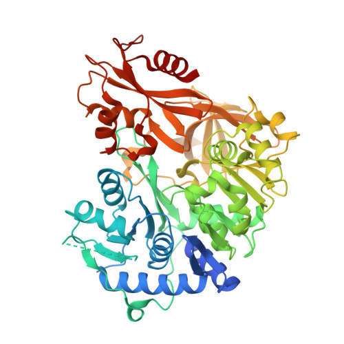
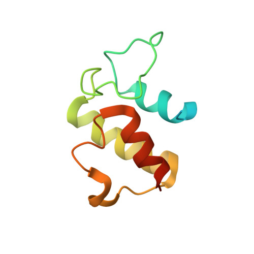
    8K4R

  • PubMed Abstract: 

    Adenylation enzymes activate amino acid substrates to aminoacyl adenylates and generally transfer this moiety onto the thiol group of the phosphopantetheine arm of a carrier protein for the selective incorporation of aminoacyl building blocks in natural product biosynthesis. In contrast to the canonical thioester-forming adenylation enzymes, the amide-forming adenylation enzyme VinM transfers an l-alanyl group onto the amino group of the aminoacyl unit attached to the phosphopantetheine arm of the carrier protein VinL to generate dipeptidyl-VinL in vicenistatin biosynthesis. It is unclear how VinM distinguishes aminoacyl-VinL from VinL for amide bond formation. Herein we describe structural and biochemical analyses of VinM. We determined the crystal structure of VinM in complex with VinL using a designed pantetheine-type cross-linking probe. The VinM-VinL complex structure in combination with site-directed mutagenesis analysis revealed that the interactions with both the phosphopantetheine arm and VinL are critical for the amide-forming activity of VinM.


  • Organizational Affiliation

    Department of Chemistry, Tokyo Institute of Technology, Tokyo 152-8551, Japan.


Macromolecules
Find similar proteins by:  (by identity cutoff)  |  3D Structure
Entity ID: 1
MoleculeChains Sequence LengthOrganismDetailsImage
Non-ribosomal peptide synthetase
A, C
540Streptomyces halstediiMutation(s): 1 
Gene Names: vinM
UniProt
Find proteins for Q76KY3 (Streptomyces halstedii)
Explore Q76KY3 
Go to UniProtKB:  Q76KY3
Entity Groups  
Sequence Clusters30% Identity50% Identity70% Identity90% Identity95% Identity100% Identity
UniProt GroupQ76KY3
Sequence Annotations
Expand
  • Reference Sequence
Find similar proteins by:  (by identity cutoff)  |  3D Structure
Entity ID: 2
MoleculeChains Sequence LengthOrganismDetailsImage
Acyl-carrier-protein
B, D
102Streptomyces halstediiMutation(s): 0 
Gene Names: vinL
UniProt
Find proteins for Q76KY4 (Streptomyces halstedii)
Explore Q76KY4 
Go to UniProtKB:  Q76KY4
Entity Groups  
Sequence Clusters30% Identity50% Identity70% Identity90% Identity95% Identity100% Identity
UniProt GroupQ76KY4
Sequence Annotations
Expand
  • Reference Sequence
Experimental Data & Validation

Experimental Data

  • Method: X-RAY DIFFRACTION
  • Resolution: 2.30 Å
  • R-Value Free: 0.251 
  • R-Value Work: 0.207 
  • R-Value Observed: 0.209 
  • Space Group: P 1
Unit Cell:
Length ( Å )Angle ( ˚ )
a = 56.501α = 94.18
b = 72.987β = 90.87
c = 80.209γ = 103.34
Software Package:
Software NamePurpose
REFMACrefinement
PDB_EXTRACTdata extraction
XDSdata reduction
Aimlessdata scaling
MOLREPphasing
Cootmodel building

Structure Validation

View Full Validation Report


Ligand Structure Quality Assessment 


Entry History & Funding Information

Deposition Data


Funding OrganizationLocationGrant Number
Japan Society for the Promotion of Science (JSPS)Japan20H02911

Revision History  (Full details and data files)

  • Version 1.0: 2023-11-01
    Type: Initial release
  • Version 1.1: 2023-11-29
    Changes: Database references