


Structure of Mangifera Indica Epoxide hydrolase 2
Bhoite, A.S., Gupta, V.S., Kulkarni, K.A.To be published.
Experimental Data Snapshot
Starting Model: experimental
View more details
Entity ID: 1 | |||||
|---|---|---|---|---|---|
| Molecule | Chains | Sequence Length | Organism | Details | Image |
| Epoxide hydrolase-2 | 324 | Mangifera indica | Mutation(s): 0 Gene Names: EH2 | 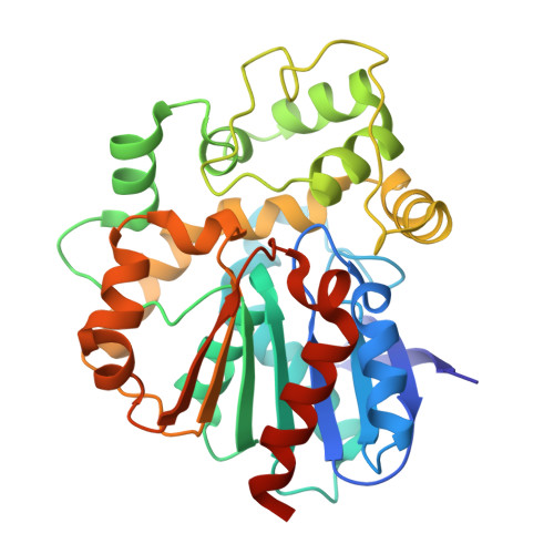 | |
UniProt | |||||
Find proteins for A0A1U9ZCB5 (Mangifera indica) Explore A0A1U9ZCB5 Go to UniProtKB: A0A1U9ZCB5 | |||||
Entity Groups | |||||
| Sequence Clusters | 30% Identity50% Identity70% Identity90% Identity95% Identity100% Identity | ||||
| UniProt Group | A0A1U9ZCB5 | ||||
Sequence AnnotationsExpand | |||||
| |||||
| Ligands 1 Unique | |||||
|---|---|---|---|---|---|
| ID | Chains | Name / Formula / InChI Key | 2D Diagram | 3D Interactions | |
| PG4 (Subject of Investigation/LOI) Query on PG4 | C [auth A] | TETRAETHYLENE GLYCOL C8 H18 O5 UWHCKJMYHZGTIT-UHFFFAOYSA-N |  | ||
| Length ( Å ) | Angle ( ˚ ) |
|---|---|
| a = 99.04 | α = 90 |
| b = 99.04 | β = 90 |
| c = 172.784 | γ = 90 |
| Software Name | Purpose |
|---|---|
| PHENIX | refinement |
| Aimless | data scaling |
| XDS | data reduction |
| PHASER | phasing |
| Funding Organization | Location | Grant Number |
|---|---|---|
| Council of Scientific & Industrial Research (CSIR) | India | -- |
RCSB PDB (citation) is hosted by
RCSB PDB is a member of the

