7TGM

Crystal structure of HSC-AMS bound DesD, the desferrioxamine synthetase from the Streptomyces griseoflavus ferrimycin biosynthetic pathway


Experimental Data Snapshot

  • Method: X-RAY DIFFRACTION
  • Resolution: 2.50 Å
  • R-Value Free: 0.220 
  • R-Value Work: 0.189 
  • R-Value Observed: 0.189 

wwPDB Validation   3D Report Full Report


Ligand Structure Quality Assessment 


This is version 1.2 of the entry. See complete history


Literature

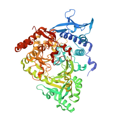
An acyl-adenylate mimic reveals the structural basis for substrate recognition by the iterative siderophore synthetase DesD.

Yang, J.Banas, V.S.Patel, K.D.Rivera, G.S.M.Mydy, L.S.Gulick, A.M.Wencewicz, T.A.

(2022) J Biol Chem 298: 102166-102166

  • DOI: https://doi.org/10.1016/j.jbc.2022.102166
  • Primary Citation of Related Structures:  
    7TGJ, 7TGK, 7TGL, 7TGM, 7TGN

  • PubMed Abstract: 

    Siderophores are conditionally essential metabolites used by microbes for environmental iron sequestration. Most Streptomyces strains produce hydroxamate-based desferrioxamine (DFO) siderophores composed of repeating units of N 1 -hydroxy-cadaverine (or N 1 -hydroxy-putrescine) and succinate. The DFO biosynthetic operon, desABCD, is highly conserved in Streptomyces; however, expression of desABCD alone does not account for the vast structural diversity within this natural product class. Here, we report the in vitro reconstitution and biochemical characterization of four DesD orthologs from Streptomyces strains that produce unique DFO siderophores. Under in vitro conditions, all four DesD orthologs displayed similar saturation steady-state kinetics (V max  = 0.9-2.5 μM⋅min -1 ) and produced the macrocyclic trimer DFOE as the favored product, suggesting a conserved role for DesD in the biosynthesis of DFO siderophores. We further synthesized a structural mimic of N 1 -hydroxy-N 1 -succinyl-cadaverine (HSC)-acyl-adenylate, the HSC-acyl sulfamoyl adenosine analog (HSC-AMS), and obtained crystal structures of DesD in the ATP-bound, AMP/PP i -bound, and HSC-AMS/P i -bound forms. We found HSC-AMS inhibited DesD orthologs (IC 50 values = 48-53 μM) leading to accumulation of linear trimeric DFOG and di-HSC at the expense of macrocyclic DFOE. Addition of exogenous PP i enhanced DesD inhibition by HSC-AMS, presumably via stabilization of the DesD-HSC-AMS complex, similar to the proposed mode of adenylate stabilization where PP i remains buried in the active site. In conclusion, our data suggest that acyl-AMS derivatives may have utility as chemical probes and bisubstrate inhibitors to reveal valuable mechanistic and structural insight for this unique family of adenylating enzymes.


  • Organizational Affiliation

    Department of Chemistry, Washington University in St Louis, St Louis, Missouri, USA.


Macromolecules
Find similar proteins by:  (by identity cutoff)  |  3D Structure
Entity ID: 1
MoleculeChains Sequence LengthOrganismDetailsImage
Desferrioxamine synthetase DesDA,
B,
C,
D [auth E],
E [auth D]
612Streptomyces griseoflavusMutation(s): 0 
Entity Groups  
Sequence Clusters30% Identity50% Identity70% Identity90% Identity95% Identity100% Identity
Sequence Annotations
Expand
  • Reference Sequence
Small Molecules
Ligands 6 Unique
IDChains Name / Formula / InChI Key2D Diagram3D Interactions
LMS (Subject of Investigation/LOI)
Query on LMS

Download Ideal Coordinates CCD File 
OC [auth E],
OD [auth D],
SB [auth C],
T [auth A],
TA [auth B]
[(2R,3S,4R,5R)-5-(6-AMINO-9H-PURIN-9-YL)-3,4-DIHYDROXYTETRAHYDRO-2-FURANYL]METHYL SULFAMATE
C10 H14 N6 O6 S
GNZLUJQJDPRUTD-KQYNXXCUSA-N
I3U (Subject of Investigation/LOI)
Query on I3U

Download Ideal Coordinates CCD File 
NC [auth E],
ND [auth D],
RB [auth C],
S [auth A],
SA [auth B]
4-[(5-aminopentyl)(hydroxy)amino]-4-oxobutanoic acid
C9 H18 N2 O4
VUXMGAKZQBQIAH-UHFFFAOYSA-N
SO4
Query on SO4

Download Ideal Coordinates CCD File 
AA [auth A]
AB [auth B]
AC [auth C]
AE [auth D]
BA [auth A]
AA [auth A],
AB [auth B],
AC [auth C],
AE [auth D],
BA [auth A],
BB [auth B],
BC [auth C],
CA [auth A],
CB [auth B],
CC [auth C],
DA [auth A],
DB [auth B],
DC [auth C],
EB [auth B],
EC [auth C],
FB [auth B],
GB [auth B],
HB [auth B],
RC [auth E],
RD [auth D],
SC [auth E],
SD [auth D],
TC [auth E],
TD [auth D],
UC [auth E],
UD [auth D],
VB [auth C],
VC [auth E],
VD [auth D],
W [auth A],
WA [auth B],
WB [auth C],
WC [auth E],
WD [auth D],
X [auth A],
XA [auth B],
XB [auth C],
XC [auth E],
XD [auth D],
Y [auth A],
YA [auth B],
YB [auth C],
YD [auth D],
Z [auth A],
ZA [auth B],
ZB [auth C],
ZD [auth D]
SULFATE ION
O4 S
QAOWNCQODCNURD-UHFFFAOYSA-L
PO4
Query on PO4

Download Ideal Coordinates CCD File 
PC [auth E],
PD [auth D],
TB [auth C],
U [auth A],
UA [auth B]
PHOSPHATE ION
O4 P
NBIIXXVUZAFLBC-UHFFFAOYSA-K
GOL
Query on GOL

Download Ideal Coordinates CCD File 
AD [auth D]
BD [auth D]
CD [auth D]
DD [auth D]
EA [auth B]
AD [auth D],
BD [auth D],
CD [auth D],
DD [auth D],
EA [auth B],
ED [auth D],
F [auth A],
FA [auth B],
FC [auth E],
FD [auth D],
G [auth A],
GA [auth B],
GC [auth E],
GD [auth D],
H [auth A],
HA [auth B],
HC [auth E],
HD [auth D],
I [auth A],
IA [auth B],
IB [auth C],
IC [auth E],
ID [auth D],
J [auth A],
JA [auth B],
JB [auth C],
JC [auth E],
JD [auth D],
K [auth A],
KA [auth B],
KB [auth C],
KC [auth E],
KD [auth D],
L [auth A],
LA [auth B],
LB [auth C],
LC [auth E],
LD [auth D],
M [auth A],
MA [auth B],
MB [auth C],
MC [auth E],
MD [auth D],
N [auth A],
NA [auth B],
NB [auth C],
O [auth A],
OA [auth B],
OB [auth C],
P [auth A],
PA [auth B],
PB [auth C],
Q [auth A],
QA [auth B],
QB [auth C],
R [auth A],
RA [auth B],
YC [auth D],
ZC [auth D]
GLYCEROL
C3 H8 O3
PEDCQBHIVMGVHV-UHFFFAOYSA-N
MG
Query on MG

Download Ideal Coordinates CCD File 
QC [auth E],
QD [auth D],
UB [auth C],
V [auth A],
VA [auth B]
MAGNESIUM ION
Mg
JLVVSXFLKOJNIY-UHFFFAOYSA-N
Experimental Data & Validation

Experimental Data

  • Method: X-RAY DIFFRACTION
  • Resolution: 2.50 Å
  • R-Value Free: 0.220 
  • R-Value Work: 0.189 
  • R-Value Observed: 0.189 
  • Space Group: C 2 2 21
Unit Cell:
Length ( Å )Angle ( ˚ )
a = 125.8α = 90
b = 237.4β = 90
c = 329.7γ = 90
Software Package:
Software NamePurpose
PHENIXrefinement
XDSdata reduction
Aimlessdata scaling
PHENIXphasing

Structure Validation

View Full Validation Report


Ligand Structure Quality Assessment 


Entry History & Funding Information

Deposition Data


Funding OrganizationLocationGrant Number
National Institutes of Health/National Institute of General Medical Sciences (NIH/NIGMS)United StatesGM136235
National Science Foundation (NSF, United States)United States1654611

Revision History  (Full details and data files)

  • Version 1.0: 2022-07-06
    Type: Initial release
  • Version 1.1: 2022-08-17
    Changes: Database references
  • Version 1.2: 2023-10-18
    Changes: Data collection, Refinement description