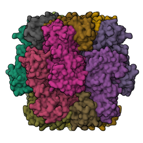
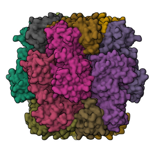
7JN4

Rubisco in the apo state

  • Classification: PLANT PROTEIN
  • Organism(s): Chlamydomonas reinhardtii
  • Mutation(s): No 

  • Deposited: 2020-08-03 Released: 2020-11-18 
  • Deposition Author(s): Matthies, D., Jonikas, M.C., He, S.
  • Funding Organization(s): National Science Foundation (NSF, United States), National Institutes of Health/National Institute of General Medical Sciences (NIH/NIGMS), Howard Hughes Medical Institute (HHMI), Ministry of Education (MoE, Singapore), Biotechnology and Biological Sciences Research Council (BBSRC), Leverhulme Trust

Experimental Data Snapshot

  • Method: ELECTRON MICROSCOPY
  • Resolution: 2.68 Å
  • Aggregation State: PARTICLE 
  • Reconstruction Method: SINGLE PARTICLE 

Starting Model: experimental
View more details

wwPDB Validation   3D Report Full Report


This is version 1.3 of the entry. See complete history


Literature

The structural basis of Rubisco phase separation in the pyrenoid.

He, S.Chou, H.T.Matthies, D.Wunder, T.Meyer, M.T.Atkinson, N.Martinez-Sanchez, A.Jeffrey, P.D.Port, S.A.Patena, W.He, G.Chen, V.K.Hughson, F.M.McCormick, A.J.Mueller-Cajar, O.Engel, B.D.Yu, Z.Jonikas, M.C.

(2020) Nat Plants 6: 1480-1490

  • DOI: https://doi.org/10.1038/s41477-020-00811-y
  • Primary Citation of Related Structures:  
    7JFO, 7JN4, 7JSX

  • PubMed Abstract: 

    Approximately one-third of global CO 2 fixation occurs in a phase-separated algal organelle called the pyrenoid. The existing data suggest that the pyrenoid forms by the phase separation of the CO 2 -fixing enzyme Rubisco with a linker protein; however, the molecular interactions underlying this phase separation remain unknown. Here we present the structural basis of the interactions between Rubisco and its intrinsically disordered linker protein Essential Pyrenoid Component 1 (EPYC1) in the model alga Chlamydomonas reinhardtii. We find that EPYC1 consists of five evenly spaced Rubisco-binding regions that share sequence similarity. Single-particle cryo-electron microscopy of these regions in complex with Rubisco indicates that each Rubisco holoenzyme has eight binding sites for EPYC1, one on each Rubisco small subunit. Interface mutations disrupt binding, phase separation and pyrenoid formation. Cryo-electron tomography supports a model in which EPYC1 and Rubisco form a codependent multivalent network of specific low-affinity bonds, giving the matrix liquid-like properties. Our results advance the structural and functional understanding of the phase separation underlying the pyrenoid, an organelle that plays a fundamental role in the global carbon cycle.


  • Organizational Affiliation

    Department of Molecular Biology, Princeton University, Princeton, NJ, USA.


Macromolecules
Find similar proteins by:  (by identity cutoff)  |  3D Structure
Entity ID: 1
MoleculeChains Sequence LengthOrganismDetailsImage
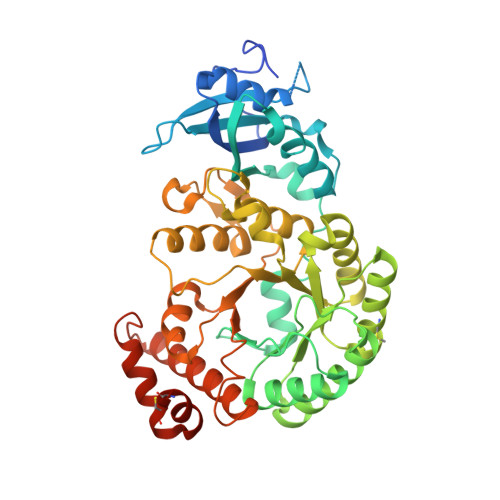
Ribulose bisphosphate carboxylase large chain
A, C, E, G, I
A, C, E, G, I, K, M, O
475Chlamydomonas reinhardtiiMutation(s): 0 
EC: 4.1.1.39
UniProt
Find proteins for P00877 (Chlamydomonas reinhardtii)
Explore P00877 
Go to UniProtKB:  P00877
Entity Groups  
Sequence Clusters30% Identity50% Identity70% Identity90% Identity95% Identity100% Identity
UniProt GroupP00877
Sequence Annotations
Expand
  • Reference Sequence
Find similar proteins by:  (by identity cutoff)  |  3D Structure
Entity ID: 2
MoleculeChains Sequence LengthOrganismDetailsImage
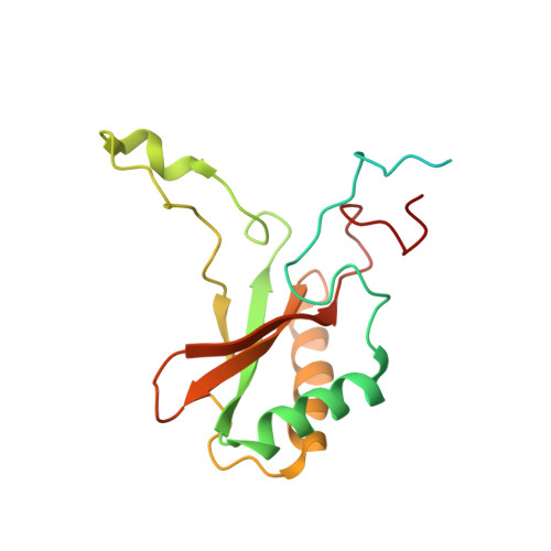
Ribulose bisphosphate carboxylase small chain 2, chloroplastic
B, D, F, H, J
B, D, F, H, J, L, N, P
185Chlamydomonas reinhardtiiMutation(s): 0 
EC: 4.1.1.39
UniProt
Find proteins for P08475 (Chlamydomonas reinhardtii)
Explore P08475 
Go to UniProtKB:  P08475
Entity Groups  
Sequence Clusters30% Identity50% Identity70% Identity90% Identity95% Identity100% Identity
UniProt GroupP08475
Sequence Annotations
Expand
  • Reference Sequence
Small Molecules
Modified Residues  1 Unique
IDChains TypeFormula2D DiagramParent
SMC
Query on SMC
A, C, E, G, I
A, C, E, G, I, K, M, O
L-PEPTIDE LINKINGC4 H9 N O2 SCYS
Experimental Data & Validation

Experimental Data

  • Method: ELECTRON MICROSCOPY
  • Resolution: 2.68 Å
  • Aggregation State: PARTICLE 
  • Reconstruction Method: SINGLE PARTICLE 
EM Software:
TaskSoftware PackageVersion
RECONSTRUCTIONRELION
MODEL REFINEMENTCoot
MODEL REFINEMENTPHENIX

Structure Validation

View Full Validation Report


Entry History & Funding Information

Deposition Data


Funding OrganizationLocationGrant Number
National Science Foundation (NSF, United States)United StatesIOS-1359682
National Science Foundation (NSF, United States)United StatesMCB-1935444
National Institutes of Health/National Institute of General Medical Sciences (NIH/NIGMS)United StatesDP2-GM-119137
Howard Hughes Medical Institute (HHMI)United States55108535
Ministry of Education (MoE, Singapore)SingaporeMOE2018-T2-2-059
Biotechnology and Biological Sciences Research Council (BBSRC)United KingdomBB/S015531/1
Leverhulme TrustUnited KingdomRPG-2017-402
National Institutes of Health/National Institute of General Medical Sciences (NIH/NIGMS)United StatesR01GM071574
National Institutes of Health/National Institute of General Medical Sciences (NIH/NIGMS)United StatesT32GM007276

Revision History  (Full details and data files)

  • Version 1.0: 2020-11-18
    Type: Initial release
  • Version 1.1: 2020-12-09
    Changes: Database references
  • Version 1.2: 2020-12-23
    Changes: Database references
  • Version 1.3: 2024-10-23
    Changes: Data collection, Database references, Refinement description, Structure summary