Molecular Insights into How the Dimetal Center in Dihydropyrimidinase Can Bind the Thymine Antagonist 5-Aminouracil: A Different Binding Mode from the Anticancer Drug 5-Fluorouracil.
Lin, E.S., Luo, R.H., Yang, Y.C., Huang, C.Y.(2022) Bioinorg Chem Appl 2022: 1817745-1817745
- PubMed: 35198016
- DOI: https://doi.org/10.1155/2022/1817745
- Primary Citation of Related Structures:
7E3U - PubMed Abstract:
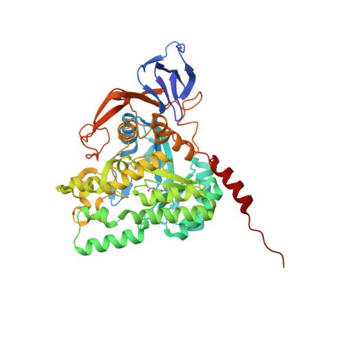
Dihydropyrimidinase (DHPase) is a key enzyme for pyrimidine degradation. DHPase contains a binuclear metal center in which two Zn ions are bridged by a posttranslationally carbamylated lysine. DHPase catalyzes the hydrolysis of dihydrouracil to N -carbamoyl- β -alanine. Whether 5-aminouracil (5-AU), a thymine antagonist and an anticancer drug that can block DNA synthesis and induce replication stress, can interact with DHPase remains to be investigated. In this study, we determined the crystal structure of Pseudomonas aeruginosa DHPase (PaDHPase) complexed with 5-AU at 2.1 Å resolution (PDB entry 7E3U). This complexed structure revealed that 5-AU interacts with Zn α (3.2 Å), Zn β (3.0 Å), the main chains of residues Ser289 (2.8 Å) and Asn337 (3.3 Å), and the side chain of residue Tyr155 (2.8 Å). These residues are also known as the substrate-binding sites of DHPase. Dynamic loop I (amino acid residues Pro65-Val70) in PaDHPase is not involved in the binding of 5-AU. The fluorescence quenching analysis and site-directed mutagenesis were used to confirm the binding mode revealed by the complexed crystal structure. The 5-AU binding mode of PaDHPase is, however, different from that of 5-fluorouracil, the best-known fluoropyrimidine used for anticancer therapy. These results provide molecular insights that may facilitate the development of new inhibitors targeting DHPase and constitute the 5-AU interactome.
Organizational Affiliation:
Department of Beauty Science, National Taichung University of Science and Technology, Taichung City, Taiwan.