6P1J | pdb_00006p1j

The structure of condensation and adenylation domains of teixobactin-producing nonribosomal peptide synthetase Txo2 serine module


Experimental Data Snapshot

  • Method: X-RAY DIFFRACTION
  • Resolution: 2.95 Å
  • R-Value Free: 
    0.267 (Depositor), 0.270 (DCC) 
  • R-Value Work: 
    0.226 (Depositor), 0.230 (DCC) 
  • R-Value Observed: 
    0.228 (Depositor) 

Starting Model: experimental
View more details

wwPDB Validation   3D Report Full Report


This is version 1.4 of the entry. See complete history


Literature

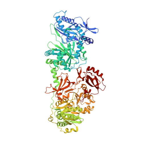
Structures of teixobactin-producing nonribosomal peptide synthetase condensation and adenylation domains.

Tan, K.Zhou, M.Jedrzejczak, R.P.Wu, R.Higuera, R.A.Borek, D.Babnigg, G.Joachimiak, A.

(2020) Curr Res Struct Biol 2: 14-24

  • DOI: https://doi.org/10.1016/j.crstbi.2020.01.002
  • Primary Citation of Related Structures:  
    6OYF, 6OZV, 6P1J, 6P3I, 6P4U

  • PubMed Abstract: 

    The recently discovered antibiotic teixobactin is produced by uncultured soil bacteria. The antibiotic inhibits cell wall synthesis of Gram-positive bacteria by binding to precursors of cell wall building blocks, and therefore it is thought to be less vulnerable to development of resistance. Teixobactin is synthesized by two nonribosomal peptide synthetases (NRPSs), encoded by txo1 and txo2 genes. Like other NRPSs, the Txo1 and Txo2 synthetases are large, multifunctional, and comprised of several modules. Each module is responsible for catalysis of a distinct step of teixobactin synthesis and contains specific functional units, commonly including a condensation (C) domain, an adenylation (A) domain, and a peptidyl carrier protein (PCP) domain. Here we report the structures of the C-A bidomains of the two L-Ser condensing modules, from Txo1 and Txo2, respectively. In the structure of the C domain of the L-Ser subunit of Txo1, a large conformational change is observed, featuring an outward swing of its N-terminal α-helix. This repositioning, if functionally validated, provides the necessary conformational change for the condensation reaction in C domain, and likely represents a regulatory mechanism. In an A core subdomain, a well-coordinated Mg 2+ cation is observed, which is required in the adenylation reaction. The Mg 2+ -binding site is defined by a largely conserved amino acid sequence motif and is coordinated by the α-phosphate group of AMP (or ATP) when present, providing some structural evidence for the role of the metal cation in the catalysis of A domain.


  • Organizational Affiliation
    • Center for Structural Genomics of Infectious Diseases, University of Chicago, 5735 South Ellis Avenue, Chicago, IL 60637, USA.

Macromolecules
Find similar proteins by:  (by identity cutoff)  |  3D Structure
Entity ID: 1
MoleculeChains Sequence LengthOrganismDetailsImage
Txo2
A, B
964Eleftheria terraeMutation(s): 0 
UniProt
Find proteins for A0A0B5H0S3 (Eleftheria terrae)
Explore A0A0B5H0S3 
Go to UniProtKB:  A0A0B5H0S3
Entity Groups  
Sequence Clusters30% Identity50% Identity70% Identity90% Identity95% Identity100% Identity
UniProt GroupA0A0B5H0S3
Sequence Annotations
Expand
  • Reference Sequence
Small Molecules
Ligands 6 Unique
IDChains Name / Formula / InChI Key2D Diagram3D Interactions
FLC
Query on FLC

Download Ideal Coordinates CCD File 
D [auth A],
W [auth B]
CITRATE ANION
C6 H5 O7
KRKNYBCHXYNGOX-UHFFFAOYSA-K
SO4
Query on SO4

Download Ideal Coordinates CCD File 
Q [auth A],
R [auth A]
SULFATE ION
O4 S
QAOWNCQODCNURD-UHFFFAOYSA-L
GOL
Query on GOL

Download Ideal Coordinates CCD File 
L [auth A],
M [auth A],
N [auth A],
O [auth A],
P [auth A]
GLYCEROL
C3 H8 O3
PEDCQBHIVMGVHV-UHFFFAOYSA-N
ACT
Query on ACT

Download Ideal Coordinates CCD File 
E [auth A]ACETATE ION
C2 H3 O2
QTBSBXVTEAMEQO-UHFFFAOYSA-M
FMT
Query on FMT

Download Ideal Coordinates CCD File 
C [auth A],
S [auth B],
T [auth B],
U [auth B],
V [auth B]
FORMIC ACID
C H2 O2
BDAGIHXWWSANSR-UHFFFAOYSA-N
CL
Query on CL

Download Ideal Coordinates CCD File 
AA [auth B]
BA [auth B]
CA [auth B]
DA [auth B]
EA [auth B]
AA [auth B],
BA [auth B],
CA [auth B],
DA [auth B],
EA [auth B],
F [auth A],
FA [auth B],
G [auth A],
GA [auth B],
H [auth A],
HA [auth B],
I [auth A],
IA [auth B],
J [auth A],
JA [auth B],
K [auth A],
X [auth B],
Y [auth B],
Z [auth B]
CHLORIDE ION
Cl
VEXZGXHMUGYJMC-UHFFFAOYSA-M
Experimental Data & Validation

Experimental Data

  • Method: X-RAY DIFFRACTION
  • Resolution: 2.95 Å
  • R-Value Free:  0.267 (Depositor), 0.270 (DCC) 
  • R-Value Work:  0.226 (Depositor), 0.230 (DCC) 
  • R-Value Observed: 0.228 (Depositor) 
Space Group: C 2 2 21
Unit Cell:
Length ( Å )Angle ( ˚ )
a = 110.857α = 90
b = 399.875β = 90
c = 144.116γ = 90
Software Package:
Software NamePurpose
PHENIXrefinement
HKL-3000data reduction
HKL-3000data scaling
HKL-3000phasing

Structure Validation

View Full Validation Report


Entry History & Funding Information

Deposition Data


Funding OrganizationLocationGrant Number
National Institutes of Health/National Institute Of Allergy and Infectious Diseases (NIH/NIAID)United StatesHHSN272201700060C

Revision History  (Full details and data files)

  • Version 1.0: 2019-05-29
    Type: Initial release
  • Version 1.1: 2019-12-18
    Changes: Author supporting evidence
  • Version 1.2: 2020-03-25
    Changes: Database references
  • Version 1.3: 2023-10-11
    Changes: Data collection, Database references, Refinement description
  • Version 1.4: 2026-03-25
    Changes: Database references, Structure summary