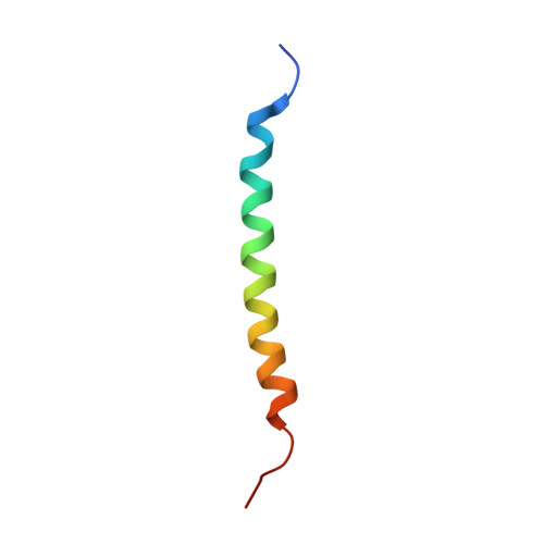
6NHW

Structure of the transmembrane domain of the Death Receptor 5 - Dimer of Trimer


Experimental Data Snapshot

  • Method: SOLUTION NMR
  • Conformers Calculated: 100 
  • Conformers Submitted: 15 
  • Selection Criteria: structures with the lowest energy 

wwPDB Validation   3D Report Full Report


This is version 1.4 of the entry. See complete history


Literature

Higher-Order Clustering of the Transmembrane Anchor of DR5 Drives Signaling.

Pan, L.Fu, T.M.Zhao, W.Zhao, L.Chen, W.Qiu, C.Liu, W.Liu, Z.Piai, A.Fu, Q.Chen, S.Wu, H.Chou, J.J.

(2019) Cell 176: 1477-1489.e14

  • DOI: https://doi.org/10.1016/j.cell.2019.02.001
  • Primary Citation of Related Structures:  
    6NHW, 6NHY

  • PubMed Abstract: 

    Receptor clustering on the cell membrane is critical in the signaling of many immunoreceptors, and this mechanism has previously been attributed to the extracellular and/or the intracellular interactions. Here, we report an unexpected finding that for death receptor 5 (DR5), a receptor in the tumor necrosis factor receptor superfamily, the transmembrane helix (TMH) alone in the receptor directly assembles a higher-order structure to drive signaling and that this structure is inhibited by the unliganded ectodomain. Nuclear magnetic resonance structure of the TMH in bicelles shows distinct trimerization and dimerization faces, allowing formation of dimer-trimer interaction networks. Single-TMH mutations that disrupt either trimerization or dimerization abolish ligand-induced receptor activation. Surprisingly, proteolytic removal of the DR5 ectodomain can fully activate downstream signaling in the absence of ligand. Our data suggest a receptor activation mechanism in which binding of ligand or antibodies to overcome the pre-ligand autoinhibition allows TMH clustering and thus signaling.


  • Organizational Affiliation

    Department of Biological Chemistry and Molecular Pharmacology, Blavatnik Institute at Harvard Medical School, 250 Longwood Avenue, Boston, MA 02115, USA.


Macromolecules
Find similar proteins by:  (by identity cutoff)  |  3D Structure
Entity ID: 1
MoleculeChains Sequence LengthOrganismDetailsImage
Tumor necrosis factor receptor superfamily member 10B
A, B, C, D, E
A, B, C, D, E, F
36Homo sapiensMutation(s): 0 
Gene Names: TNFRSF10BDR5KILLERTRAILR2TRICK2ZTNFR9UNQ160/PRO186
Membrane Entity: Yes 
UniProt & NIH Common Fund Data Resources
Find proteins for O14763 (Homo sapiens)
Explore O14763 
Go to UniProtKB:  O14763
PHAROS:  O14763
GTEx:  ENSG00000120889 
Entity Groups  
Sequence Clusters30% Identity50% Identity70% Identity90% Identity95% Identity100% Identity
UniProt GroupO14763
Sequence Annotations
Expand
  • Reference Sequence
Experimental Data & Validation

Experimental Data

  • Method: SOLUTION NMR
  • Conformers Calculated: 100 
  • Conformers Submitted: 15 
  • Selection Criteria: structures with the lowest energy 

Structure Validation

View Full Validation Report


Entry History & Funding Information

Deposition Data


Funding OrganizationLocationGrant Number
National Institutes of Health/National Institute of General Medical Sciences (NIH/NIGMS)United StatesGM116898

Revision History  (Full details and data files)

  • Version 1.0: 2019-02-27
    Type: Initial release
  • Version 1.1: 2019-03-20
    Changes: Data collection, Database references
  • Version 1.2: 2020-01-01
    Changes: Author supporting evidence, Data collection
  • Version 1.3: 2023-06-14
    Changes: Database references, Other
  • Version 1.4: 2024-05-15
    Changes: Data collection, Database references