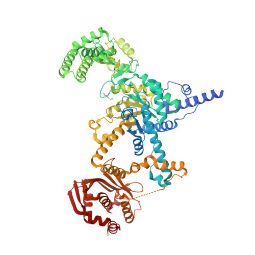
6EDZ

Crystal structure of Mycobacterium tuberculosis ICL2 in complex with acetyl-CoA, form I


Experimental Data Snapshot

  • Method: X-RAY DIFFRACTION
  • Resolution: 2.67 Å
  • R-Value Free: 0.241 
  • R-Value Work: 0.219 
  • R-Value Observed: 0.220 

wwPDB Validation   3D Report Full Report


Ligand Structure Quality Assessment 

Created with Raphaël 2.3.0Worse 01 BetterLigand structure goodness of fit to experimental dataBest fitted ACOClick on this verticalbar to view details

This is version 1.1 of the entry. See complete history


Literature

Acetyl-CoA-mediated activation of Mycobacterium tuberculosis isocitrate lyase 2.

Bhusal, R.P.Jiao, W.Kwai, B.X.C.Reynisson, J.Collins, A.J.Sperry, J.Bashiri, G.Leung, I.K.H.

(2019) Nat Commun 10: 4639-4639

  • DOI: https://doi.org/10.1038/s41467-019-12614-7
  • Primary Citation of Related Structures:  
    6EDW, 6EDZ, 6EE1

  • PubMed Abstract: 

    Isocitrate lyase is important for lipid utilisation by Mycobacterium tuberculosis but its ICL2 isoform is poorly understood. Here we report that binding of the lipid metabolites acetyl-CoA or propionyl-CoA to ICL2 induces a striking structural rearrangement, substantially increasing isocitrate lyase and methylisocitrate lyase activities. Thus, ICL2 plays a pivotal role regulating carbon flux between the tricarboxylic acid (TCA) cycle, glyoxylate shunt and methylcitrate cycle at high lipid concentrations, a mechanism essential for bacterial growth and virulence.


  • Organizational Affiliation

    School of Chemical Sciences, The University of Auckland, Private Bag 92019, Victoria Street West, Auckland, 1142, New Zealand.


Macromolecules
Find similar proteins by:  (by identity cutoff)  |  3D Structure
Entity ID: 1
MoleculeChains Sequence LengthOrganismDetailsImage
Isocitrate lyase 2
A, B, C, D
786Mycobacterium tuberculosis CDC1551Mutation(s): 0 
Gene Names: icl2aceA-2MT1966
EC: 4.1.3.1
UniProt
Find proteins for Q8VJU4 (Mycobacterium tuberculosis (strain CDC 1551 / Oshkosh))
Explore Q8VJU4 
Go to UniProtKB:  Q8VJU4
Entity Groups  
Sequence Clusters30% Identity50% Identity70% Identity90% Identity95% Identity100% Identity
UniProt GroupQ8VJU4
Sequence Annotations
Expand
  • Reference Sequence
Experimental Data & Validation

Experimental Data

  • Method: X-RAY DIFFRACTION
  • Resolution: 2.67 Å
  • R-Value Free: 0.241 
  • R-Value Work: 0.219 
  • R-Value Observed: 0.220 
  • Space Group: P 1 21 1
Unit Cell:
Length ( Å )Angle ( ˚ )
a = 105.423α = 90
b = 170.741β = 95
c = 107.011γ = 90
Software Package:
Software NamePurpose
REFMACrefinement
XDSdata reduction
Aimlessdata scaling
PHASERphasing

Structure Validation

View Full Validation Report


Ligand Structure Quality Assessment 

Created with Raphaël 2.3.0Worse 01 BetterLigand structure goodness of fit to experimental dataBest fitted ACOClick on this verticalbar to view details

Entry History 

Deposition Data

Revision History  (Full details and data files)

  • Version 1.0: 2019-08-14
    Type: Initial release
  • Version 1.1: 2019-10-23
    Changes: Data collection, Database references