6CN7

The structure of aerobactin synthetase IucC from a hypervirulent pathotype of Klebsiella pneumoniae

  • Classification: LIGASE
  • Organism(s): Klebsiella pneumoniae
  • Expression System: Escherichia coli
  • Mutation(s): Yes 

  • Deposited: 2018-03-07 Released: 2018-04-11 
  • Deposition Author(s): Bailey, D.C., Rice, M.R., Gulick, A.M.
  • Funding Organization(s): National Institutes of Health/National Institute Of Allergy and Infectious Diseases (NIH/NIAID), National Institutes of Health/National Center for Advancing Translational Sciences (NIH/NCATS)

Experimental Data Snapshot

  • Method: X-RAY DIFFRACTION
  • Resolution: 2.45 Å
  • R-Value Free: 0.240 
  • R-Value Work: 0.213 
  • R-Value Observed: 0.214 

wwPDB Validation   3D Report Full Report


This is version 1.4 of the entry. See complete history


Literature

Structural and functional delineation of aerobactin biosynthesis in hypervirulentKlebsiella pneumoniae.

Bailey, D.C.Alexander, E.Rice, M.R.Drake, E.J.Mydy, L.S.Aldrich, C.C.Gulick, A.M.

(2018) J Biol Chem 293: 7841-7852

  • DOI: https://doi.org/10.1074/jbc.RA118.002798
  • Primary Citation of Related Structures:  
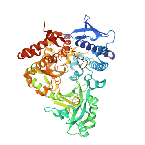
    6CN7

  • PubMed Abstract: 

    Aerobactin, a citryl-hydroxamate siderophore, is produced by a number of pathogenic Gram-negative bacteria to aid in iron assimilation. Interest in this well-known siderophore was reignited by recent investigations suggesting that it plays a key role in mediating the enhanced virulence of a hypervirulent pathotype of Klebsiella pneumoniae (hvKP). In contrast to classical opportunistic strains of K. pneumoniae , hvKP causes serious life-threatening infections in previously healthy individuals in the community. Multiple contemporary reports have confirmed fears that the convergence of multidrug-resistant and hvKP pathotypes has led to the evolution of a highly transmissible, drug-resistant, and virulent "super bug." Despite hvKP harboring four distinct siderophore operons, knocking out production of only aerobactin led to a significant attenuation of virulence. Herein, we continue our structural and functional studies on the biosynthesis of this crucial virulence factor. In vivo heterologous production and in vitro reconstitution of aerobactin biosynthesis from hvKP was carried out, demonstrating the specificity, stereoselectivity, and kinetic throughput of the complete pathway. Additionally, we present a steady-state kinetic analysis and the X-ray crystal structure of the second aerobactin synthetase IucC, as well as describe a surface entropy reduction strategy that was employed for structure determination. Finally, we show solution X-ray scattering data that support a unique dimeric quaternary structure for IucC. These new insights into aerobactin assembly will help inform potential antivirulence strategies and advance our understanding of siderophore biosynthesis.


  • Organizational Affiliation

    From the Department of Structural Biology, The Jacobs School of Medicine & Biomedical Sciences, State University of New York, Buffalo, New York 14203.


Macromolecules
Find similar proteins by:  (by identity cutoff)  |  3D Structure
Entity ID: 1
MoleculeChains Sequence LengthOrganismDetailsImage
Aerobactin synthase IucC
A, B, C, D, E
A, B, C, D, E, F, G, H
579Klebsiella pneumoniaeMutation(s): 5 
Gene Names: iucCBCB67_13855LV109
UniProt
Find proteins for Q6U605 (Klebsiella pneumoniae CG43)
Explore Q6U605 
Go to UniProtKB:  Q6U605
Entity Groups  
Sequence Clusters30% Identity50% Identity70% Identity90% Identity95% Identity100% Identity
UniProt GroupQ6U605
Sequence Annotations
Expand
  • Reference Sequence
Small Molecules
Ligands 1 Unique
IDChains Name / Formula / InChI Key2D Diagram3D Interactions
MES
Query on MES

Download Ideal Coordinates CCD File 
I [auth A],
J [auth G]
2-(N-MORPHOLINO)-ETHANESULFONIC ACID
C6 H13 N O4 S
SXGZJKUKBWWHRA-UHFFFAOYSA-N
Experimental Data & Validation

Experimental Data

  • Method: X-RAY DIFFRACTION
  • Resolution: 2.45 Å
  • R-Value Free: 0.240 
  • R-Value Work: 0.213 
  • R-Value Observed: 0.214 
  • Space Group: P 1 21 1
Unit Cell:
Length ( Å )Angle ( ˚ )
a = 124.02α = 90
b = 197.69β = 109.6
c = 130.38γ = 90
Software Package:
Software NamePurpose
PHENIXrefinement
PDB_EXTRACTdata extraction
iMOSFLMdata reduction
Cootmodel building
PHASERphasing

Structure Validation

View Full Validation Report


Entry History & Funding Information

Deposition Data


Funding OrganizationLocationGrant Number
National Institutes of Health/National Institute Of Allergy and Infectious Diseases (NIH/NIAID)United States1AI116998
National Institutes of Health/National Institute Of Allergy and Infectious Diseases (NIH/NIAID)United StatesAI007614
National Institutes of Health/National Center for Advancing Translational Sciences (NIH/NCATS)United StatesUL1TR001412

Revision History  (Full details and data files)

  • Version 1.0: 2018-04-11
    Type: Initial release
  • Version 1.1: 2018-04-18
    Changes: Data collection, Database references
  • Version 1.2: 2018-05-30
    Changes: Data collection, Database references
  • Version 1.3: 2019-12-04
    Changes: Author supporting evidence
  • Version 1.4: 2023-10-04
    Changes: Data collection, Database references, Refinement description