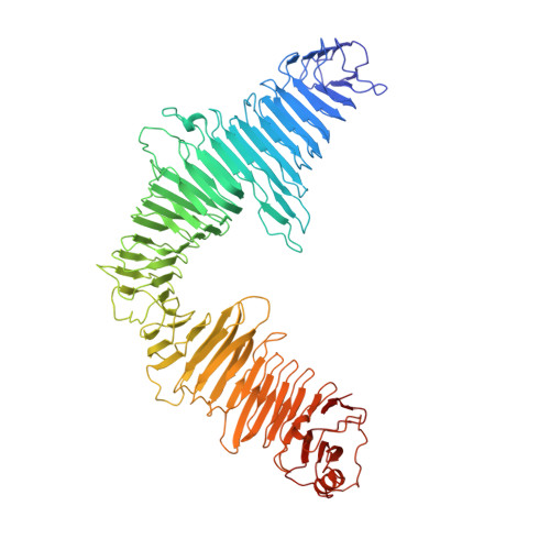
5N97

Structure of the C. crescentus S-layer


Experimental Data Snapshot

  • Method: ELECTRON MICROSCOPY
  • Resolution: 7.40 Å
  • Aggregation State: PARTICLE 
  • Reconstruction Method: SUBTOMOGRAM AVERAGING 

wwPDB Validation   3D Report Full Report


This is version 1.2 of the entry. See complete history


Literature

Structure of the hexagonal surface layer on Caulobacter crescentus cells.

Bharat, T.A.M.Kureisaite-Ciziene, D.Hardy, G.G.Yu, E.W.Devant, J.M.Hagen, W.J.H.Brun, Y.V.Briggs, J.A.G.Lowe, J.

(2017) Nat Microbiol 2: 17059-17059

  • DOI: https://doi.org/10.1038/nmicrobiol.2017.59
  • Primary Citation of Related Structures:  
    5N8P, 5N97

  • PubMed Abstract: 

    Many prokaryotic cells are encapsulated by a surface layer (S-layer) consisting of repeating units of S-layer proteins. S-layer proteins are a diverse class of molecules found in Gram-positive and Gram-negative bacteria and most archaea 1-5 . S-layers protect cells from the outside, provide mechanical stability and also play roles in pathogenicity. In situ structural information about this highly abundant class of proteins is scarce, so atomic details of how S-layers are arranged on the surface of cells have remained elusive. Here, using purified Caulobacter crescentus' sole S-layer protein RsaA, we obtained a 2.7 Å X-ray structure that shows the hexameric S-layer lattice. We also solved a 7.4 Å structure of the S-layer through electron cryotomography and sub-tomogram averaging of cell stalks. The X-ray structure was docked unambiguously into the electron cryotomography map, resulting in a pseudo-atomic-level description of the in vivo S-layer, which agrees completely with the atomic X-ray lattice model. The cellular S-layer atomic structure shows that the S-layer is porous, with a largest gap dimension of 27 Å, and is stabilized by multiple Ca 2+ ions bound near the interfaces. This study spans different spatial scales from atoms to cells by combining X-ray crystallography with electron cryotomography and sub-nanometre-resolution sub-tomogram averaging.


  • Organizational Affiliation

    Structural Studies Division, MRC Laboratory of Molecular Biology, Cambridge CB2 0QH, UK.


Macromolecules
Find similar proteins by:  (by identity cutoff)  |  3D Structure
Entity ID: 1
MoleculeChains Sequence LengthOrganismDetailsImage
S-layer protein rsaA
A, B, C, D, E
A, B, C, D, E, F
778Caulobacter vibrioides NA1000Mutation(s): 0 
UniProt
Find proteins for A0A0H3C8J1 (Caulobacter vibrioides (strain NA1000 / CB15N))
Explore A0A0H3C8J1 
Go to UniProtKB:  A0A0H3C8J1
Entity Groups  
Sequence Clusters30% Identity50% Identity70% Identity90% Identity95% Identity100% Identity
UniProt GroupA0A0H3C8J1
Sequence Annotations
Expand
  • Reference Sequence
Small Molecules
Ligands 1 Unique
IDChains Name / Formula / InChI Key2D Diagram3D Interactions
CA
Query on CA

Download Ideal Coordinates CCD File 
AA [auth B]
AB [auth C]
AC [auth D]
AD [auth F]
BA [auth B]
AA [auth B],
AB [auth C],
AC [auth D],
AD [auth F],
BA [auth B],
BB [auth C],
BC [auth D],
BD [auth F],
CA [auth B],
CB [auth C],
CC [auth D],
CD [auth F],
DA [auth B],
DB [auth C],
DC [auth D],
DD [auth F],
EA [auth B],
EB [auth C],
EC [auth E],
ED [auth F],
FA [auth B],
FB [auth C],
FC [auth E],
FD [auth F],
G [auth A],
GA [auth B],
GB [auth C],
GC [auth E],
GD [auth F],
H [auth A],
HA [auth B],
HB [auth C],
HC [auth E],
HD [auth F],
I [auth A],
IA [auth B],
IB [auth C],
IC [auth E],
ID [auth F],
J [auth A],
JA [auth B],
JB [auth C],
JC [auth E],
JD [auth F],
K [auth A],
KA [auth B],
KB [auth C],
KC [auth E],
KD [auth F],
L [auth A],
LA [auth B],
LB [auth D],
LC [auth E],
LD [auth F],
M [auth A],
MA [auth B],
MB [auth D],
MC [auth E],
MD [auth F],
N [auth A],
NA [auth B],
NB [auth D],
NC [auth E],
ND [auth F],
O [auth A],
OA [auth B],
OB [auth D],
OC [auth E],
OD [auth F],
P [auth A],
PA [auth B],
PB [auth D],
PC [auth E],
PD [auth F],
Q [auth A],
QA [auth B],
QB [auth D],
QC [auth E],
R [auth A],
RA [auth B],
RB [auth D],
RC [auth E],
S [auth A],
SA [auth C],
SB [auth D],
SC [auth E],
T [auth A],
TA [auth C],
TB [auth D],
TC [auth E],
U [auth A],
UA [auth C],
UB [auth D],
UC [auth E],
V [auth A],
VA [auth C],
VB [auth D],
VC [auth E],
W [auth A],
WA [auth C],
WB [auth D],
WC [auth E],
X [auth A],
XA [auth C],
XB [auth D],
XC [auth F],
Y [auth A],
YA [auth C],
YB [auth D],
YC [auth F],
Z [auth B],
ZA [auth C],
ZB [auth D],
ZC [auth F]
CALCIUM ION
Ca
BHPQYMZQTOCNFJ-UHFFFAOYSA-N
Experimental Data & Validation

Experimental Data

  • Method: ELECTRON MICROSCOPY
  • Resolution: 7.40 Å
  • Aggregation State: PARTICLE 
  • Reconstruction Method: SUBTOMOGRAM AVERAGING 

Structure Validation

View Full Validation Report


Entry History & Funding Information

Deposition Data


Funding OrganizationLocationGrant Number
Wellcome TrustUnited Kingdom095514/Z/11/Z
Medical Research Council (United Kingdom)United KingdomU105184326
European Molecular Biology OrganizationGermanyaALTF 778-2015

Revision History  (Full details and data files)

  • Version 1.0: 2017-04-19
    Type: Initial release
  • Version 1.1: 2017-08-02
    Changes: Data collection, Derived calculations, Refinement description
  • Version 1.2: 2018-10-24
    Changes: Data collection, Database references