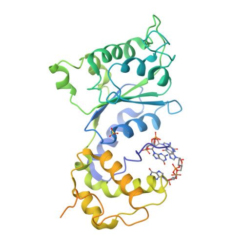
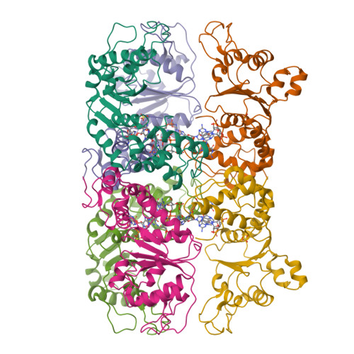
5EXX | pdb_00005exx

AAA+ ATPase FleQ from Pseudomonas aeruginosa bound to c-di-GMP


Experimental Data Snapshot

  • Method: X-RAY DIFFRACTION
  • Resolution: 3.31 Å
  • R-Value Free: 
    0.290 (Depositor), 0.290 (DCC) 
  • R-Value Work: 
    0.263 (Depositor), 0.290 (DCC) 
  • R-Value Observed: 
    0.264 (Depositor) 

wwPDB Validation   3D Report Full Report


Ligand Structure Quality Assessment 

Created with Raphaël 2.3.0Worse 01 BetterLigand structure goodness of fit to experimental dataBest fitted C2EClick on this verticalbar to view details

This is version 1.4 of the entry. See complete history


Literature

Mechanistic insights into c-di-GMP-dependent control of the biofilm regulator FleQ from Pseudomonas aeruginosa.

Matsuyama, B.Y.Krasteva, P.V.Baraquet, C.Harwood, C.S.Sondermann, H.Navarro, M.V.

(2016) Proc Natl Acad Sci U S A 113: E209-E218

  • DOI: https://doi.org/10.1073/pnas.1523148113
  • Primary Citation of Related Structures:  
    5EXP, 5EXS, 5EXT, 5EXX

  • PubMed Abstract: 

    Bacterial biofilm formation during chronic infections confers increased fitness, antibiotic tolerance, and cytotoxicity. In many pathogens, the transition from a planktonic lifestyle to collaborative, sessile biofilms represents a regulated process orchestrated by the intracellular second-messenger c-di-GMP. A main effector for c-di-GMP signaling in the opportunistic pathogen Pseudomonas aeruginosa is the transcription regulator FleQ. FleQ is a bacterial enhancer-binding protein (bEBP) with a central AAA+ ATPase σ(54)-interaction domain, flanked by a C-terminal helix-turn-helix DNA-binding motif and a divergent N-terminal receiver domain. Together with a second ATPase, FleN, FleQ regulates the expression of flagellar and exopolysaccharide biosynthesis genes in response to cellular c-di-GMP. Here we report structural and functional data that reveal an unexpected mode of c-di-GMP recognition that is associated with major conformational rearrangements in FleQ. Crystal structures of FleQ's AAA+ ATPase domain in its apo-state or bound to ADP or ATP-γ-S show conformations reminiscent of the activated ring-shaped assemblies of other bEBPs. As revealed by the structure of c-di-GMP-complexed FleQ, the second messenger interacts with the AAA+ ATPase domain at a site distinct from the ATP binding pocket. c-di-GMP interaction leads to active site obstruction, hexameric ring destabilization, and discrete quaternary structure transitions. Solution and cell-based studies confirm coupling of the ATPase active site and c-di-GMP binding, as well as the functional significance of crystallographic interprotomer interfaces. Taken together, our data offer unprecedented insight into conserved regulatory mechanisms of gene expression under direct c-di-GMP control via FleQ and FleQ-like bEBPs.


  • Organizational Affiliation

    Departamento de Física e Ciência Interdisciplinar, Instituto de Física de São Carlos, Universidade de São Paulo, São Carlos 13563-120, SP, Brazil;


Macromolecules
Find similar proteins by:  (by identity cutoff)  |  3D Structure
Entity ID: 1
MoleculeChains Sequence LengthOrganismDetailsImage
Transcriptional regulator FleQ343Pseudomonas aeruginosa PAO1Mutation(s): 0 
Gene Names: fleQPA1097
UniProt
Find proteins for G3XCV0 (Pseudomonas aeruginosa (strain ATCC 15692 / DSM 22644 / CIP 104116 / JCM 14847 / LMG 12228 / 1C / PRS 101 / PAO1))
Explore G3XCV0 
Go to UniProtKB:  G3XCV0
Entity Groups  
Sequence Clusters30% Identity50% Identity70% Identity90% Identity95% Identity100% Identity
UniProt GroupG3XCV0
Sequence Annotations
Expand
  • Reference Sequence
Small Molecules
Ligands 2 Unique
IDChains Name / Formula / InChI Key2D Diagram3D Interactions
C2E
Query on C2E

Download Ideal Coordinates CCD File 
C [auth A],
D [auth A]
9,9'-[(2R,3R,3aS,5S,7aR,9R,10R,10aS,12S,14aR)-3,5,10,12-tetrahydroxy-5,12-dioxidooctahydro-2H,7H-difuro[3,2-d:3',2'-j][1,3,7,9,2,8]tetraoxadiphosphacyclododecine-2,9-diyl]bis(2-amino-1,9-dihydro-6H-purin-6-one)
C20 H24 N10 O14 P2
PKFDLKSEZWEFGL-MHARETSRSA-N
SO4
Query on SO4

Download Ideal Coordinates CCD File 
B [auth A]SULFATE ION
O4 S
QAOWNCQODCNURD-UHFFFAOYSA-L
Experimental Data & Validation

Experimental Data

  • Method: X-RAY DIFFRACTION
  • Resolution: 3.31 Å
  • R-Value Free:  0.290 (Depositor), 0.290 (DCC) 
  • R-Value Work:  0.263 (Depositor), 0.290 (DCC) 
  • R-Value Observed: 0.264 (Depositor) 
Space Group: P 63 2 2
Unit Cell:
Length ( Å )Angle ( ˚ )
a = 137.836α = 90
b = 137.836β = 90
c = 172.773γ = 120
Software Package:
Software NamePurpose
PHENIXrefinement
XDSdata reduction
XDSdata scaling
PHENIXphasing

Structure Validation

View Full Validation Report


Ligand Structure Quality Assessment 

Created with Raphaël 2.3.0Worse 01 BetterLigand structure goodness of fit to experimental dataBest fitted C2EClick on this verticalbar to view details

Entry History & Funding Information

Deposition Data


Funding OrganizationLocationGrant Number
Sao Paulo Research Foundation (FAPESP)Brazil2009/13238-0
National Institutes of Health/National Institute Of Allergy and Infectious Diseases (NIH/NIAID)United StatesR01-AI097307

Revision History  (Full details and data files)

  • Version 1.0: 2016-02-10
    Type: Initial release
  • Version 1.1: 2017-09-13
    Changes: Author supporting evidence, Derived calculations
  • Version 1.2: 2019-04-17
    Changes: Author supporting evidence, Data collection
  • Version 1.3: 2019-12-11
    Changes: Author supporting evidence
  • Version 1.4: 2024-03-06
    Changes: Data collection, Database references, Structure summary