5BK7

The structure of MppP E15A mutant soaked with the substrate L-arginine


Experimental Data Snapshot

  • Method: X-RAY DIFFRACTION
  • Resolution: 2.20 Å
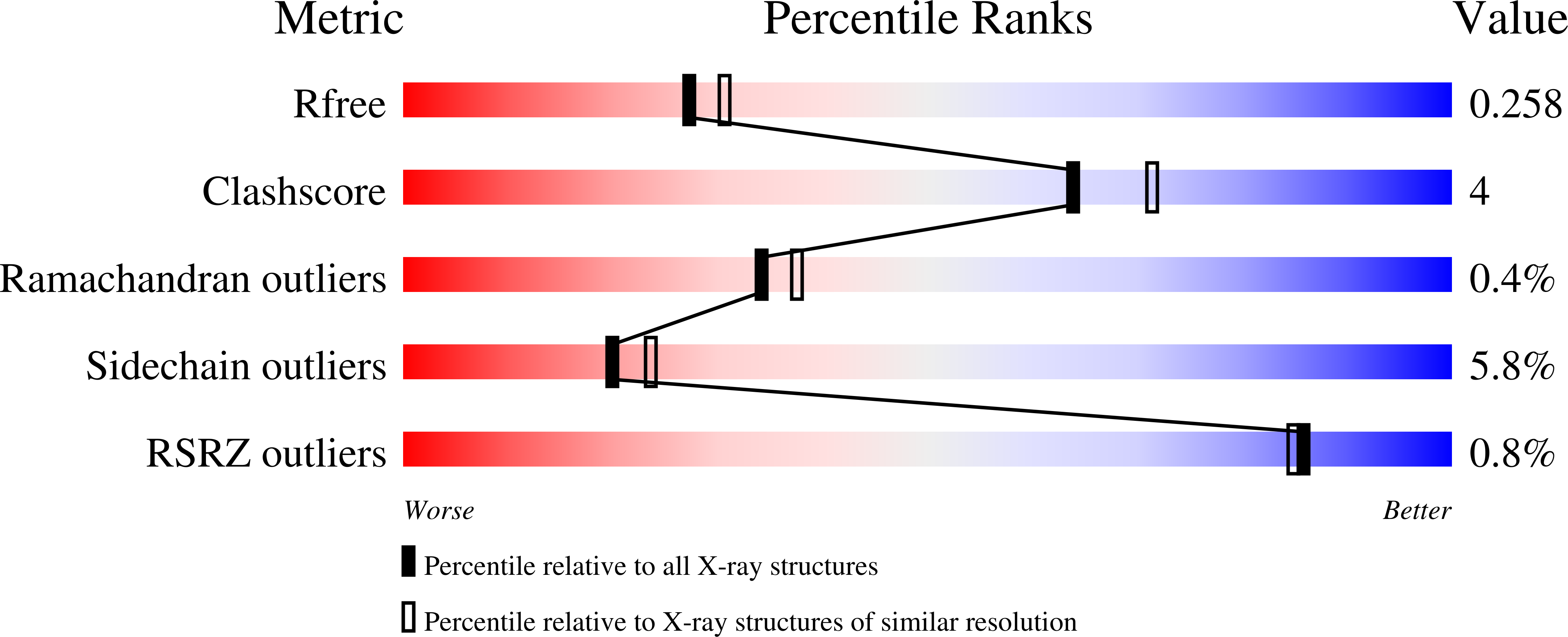
  • R-Value Free: 0.258 
  • R-Value Work: 0.201 
  • R-Value Observed: 0.202 

Starting Model: experimental
View more details

wwPDB Validation   3D Report Full Report


Ligand Structure Quality Assessment 


This is version 1.2 of the entry. See complete history


Literature

Streptomyces wadayamensis MppP is a PLP-Dependent Oxidase, Not an Oxygenase.

Han, L.Vuksanovic, N.Oehm, S.A.Fenske, T.G.Schwabacher, A.W.Silvaggi, N.R.

(2018) Biochemistry 57: 3252-3264

  • DOI: https://doi.org/10.1021/acs.biochem.8b00130
  • Primary Citation of Related Structures:  
    5BK7, 6C8T, 6C92, 6C9B

  • PubMed Abstract: 

    The PLP-dependent l-arginine hydroxylase/deaminase MppP from Streptomyces wadayamensis (SwMppP) is involved in the biosynthesis of l-enduracididine, a nonproteinogenic amino acid found in several nonribosomally produced peptide antibiotics. SwMppP uses only PLP and molecular oxygen to catalyze a 4-electron oxidation of l-arginine to form a mixture of 2-oxo-4(S)-hydroxy-5-guanidinovaleric acid and 2-oxo-5-guanidinovaleric acid. Steady-state kinetics analysis in the presence and absence of catalase shows that one molecule of peroxide is formed for every molecule of dioxygen consumed in the reaction. Moreover, for each molecule of 2-oxo-4(S)-hydroxy-5-guanidinovaleric acid produced, two molecules of dioxygen are consumed, suggesting that both the 4-hydroxy and 2-keto groups are derived from water. This was confirmed by running the reactions using either [18] O 2 or H 2 [18] O and analyzing the products by ESI-MS. Incorporation of [18] O was only observed when the reaction was performed in H 2 [18] O. Crystal structures of SwMppP with l-arginine, 2-oxo-4(S)-hydroxy-5-guanidinovaleric acid, or 2-oxo-5-guanidinovaleric acid bound were determined at resolutions of 2.2, 1.9. and 1.8 Å, respectively. The structural data show that the N-terminal portion of the protein is disordered unless substrate or product is bound in the active site, in which case it forms a well-ordered helix that covers the catalytic center. This observation suggested that the N-terminal helix may have a role in substrate binding and/or catalysis. Our structural and kinetic characterizations of N-terminal variants show that the N-terminus is critical for catalysis. In light of this new information, we have refined our previously proposed mechanism of the SwMppP-catalyzed oxidation of l-arginine.


  • Organizational Affiliation

    Department of Chemistry and Biochemistry , University of Wisconsin-Milwaukee , Milwaukee , Wisconsin 53201 , United States.


Macromolecules
Find similar proteins by:  (by identity cutoff)  |  3D Structure
Entity ID: 1
MoleculeChains Sequence LengthOrganismDetailsImage
PLP-Dependent L-Arginine Hydroxylase MppP
A, B, C, D, E
A, B, C, D, E, F, G, H
376Streptomyces wadayamensisMutation(s): 1 
UniProt
Find proteins for A0A0X1KHF5 (Streptomyces wadayamensis)
Explore A0A0X1KHF5 
Go to UniProtKB:  A0A0X1KHF5
Entity Groups  
Sequence Clusters30% Identity50% Identity70% Identity90% Identity95% Identity100% Identity
UniProt GroupA0A0X1KHF5
Sequence Annotations
Expand
  • Reference Sequence
Small Molecules
Ligands 1 Unique
IDChains Name / Formula / InChI Key2D Diagram3D Interactions
EQJ
Query on EQJ

Download Ideal Coordinates CCD File 
I [auth A]
J [auth B]
K [auth C]
L [auth D]
M [auth E]
I [auth A],
J [auth B],
K [auth C],
L [auth D],
M [auth E],
N [auth F],
O [auth G],
P [auth H]
(E)-N~2~-({3-hydroxy-2-methyl-5-[(phosphonooxy)methyl]pyridin-4-yl}methylidene)-L-arginine
C14 H22 N5 O7 P
PFHOMURYEGODJI-GBCOYWTISA-N
Experimental Data & Validation

Experimental Data

  • Method: X-RAY DIFFRACTION
  • Resolution: 2.20 Å
  • R-Value Free: 0.258 
  • R-Value Work: 0.201 
  • R-Value Observed: 0.202 
  • Space Group: P 1 21 1
Unit Cell:
Length ( Å )Angle ( ˚ )
a = 64.441α = 90
b = 200.514β = 103.25
c = 139.93γ = 90
Software Package:
Software NamePurpose
PHENIXrefinement
HKL-2000data reduction
HKL-2000data scaling
PHASERphasing

Structure Validation

View Full Validation Report


Ligand Structure Quality Assessment 


Entry History & Funding Information

Deposition Data


Funding OrganizationLocationGrant Number
National Science Foundation (NSF, United States)United StatesCHE-1606842

Revision History  (Full details and data files)

  • Version 1.0: 2018-09-05
    Type: Initial release
  • Version 1.1: 2019-11-27
    Changes: Author supporting evidence
  • Version 1.2: 2023-09-27
    Changes: Advisory, Data collection, Database references, Refinement description