4V03

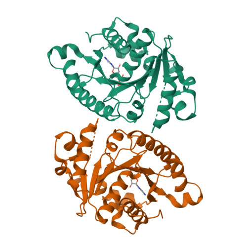
MinD cell division protein, Aquifex aeolicus


Experimental Data Snapshot

  • Method: X-RAY DIFFRACTION
  • Resolution: 1.90 Å
  • R-Value Free: 0.279 
  • R-Value Work: 0.216 
  • R-Value Observed: 0.219 

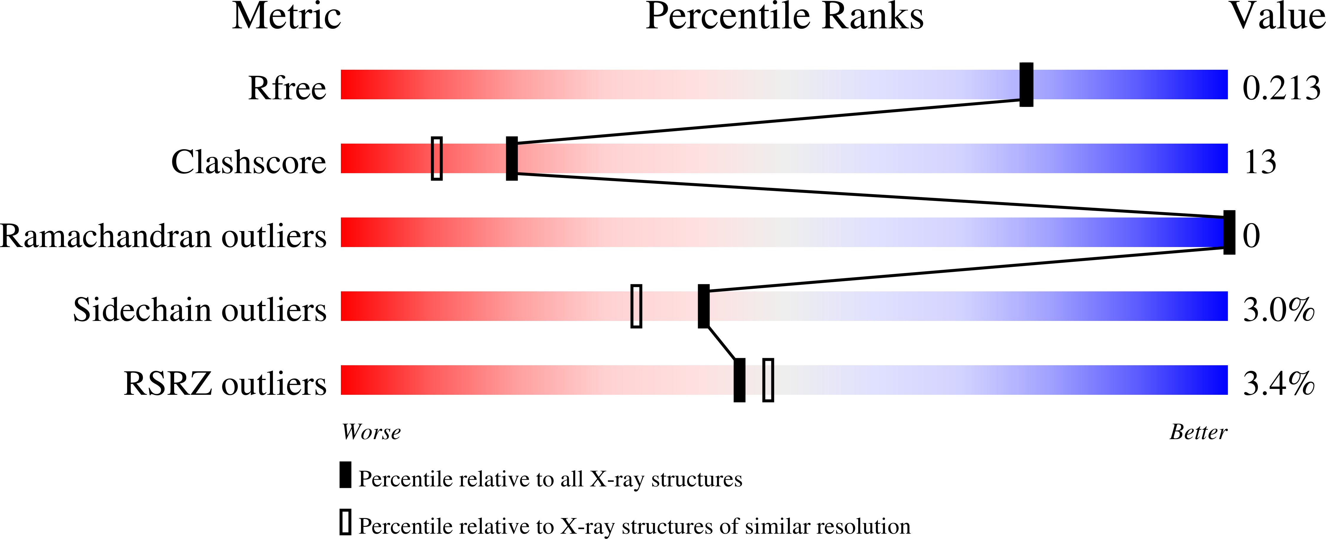
wwPDB Validation   3D Report Full Report


Ligand Structure Quality Assessment 


This is version 1.1 of the entry. See complete history


Literature

Mincd Cell Division Proteins Form Alternating Copolymeric Cytomotive Filaments.

Ghosal, D.Trambaiolo, D.Amos, L.A.Lowe, J.

(2014) Nat Commun 5: 5341

  • DOI: https://doi.org/10.1038/ncomms6341
  • Primary Citation of Related Structures:  
    4V02, 4V03

  • PubMed Abstract: 

    During bacterial cell division, filaments of the tubulin-like protein FtsZ assemble at midcell to form the cytokinetic Z-ring. Its positioning is regulated by the oscillation of MinCDE proteins. MinC is activated by MinD through an unknown mechanism and prevents Z-ring assembly anywhere but midcell. Here, using X-ray crystallography, electron microscopy and in vivo analyses, we show that MinD activates MinC by forming a new class of alternating copolymeric filaments that show similarity to eukaryotic septin filaments. A non-polymerizing mutation in MinD causes aberrant cell division in Escherichia coli. MinCD copolymers bind to membrane, interact with FtsZ and are disassembled by MinE. Imaging a functional msfGFP-MinC fusion protein in MinE-deleted cells reveals filamentous structures. EM imaging of our reconstitution of the MinCD-FtsZ interaction on liposome surfaces reveals a plausible mechanism for regulation of FtsZ ring assembly by MinCD copolymers.


  • Organizational Affiliation

    Structural Studies, MRC Laboratory of Molecular Biology, Francis Crick Avenue, Cambridge CB2 0QH, UK.


Macromolecules
Find similar proteins by:  (by identity cutoff)  |  3D Structure
Entity ID: 1
MoleculeChains Sequence LengthOrganismDetailsImage
SITE-DETERMINING PROTEIN
A, B
257Aquifex aeolicusMutation(s): 0 
UniProt
Find proteins for O67033 (Aquifex aeolicus (strain VF5))
Explore O67033 
Go to UniProtKB:  O67033
Entity Groups  
Sequence Clusters30% Identity50% Identity70% Identity90% Identity95% Identity100% Identity
UniProt GroupO67033
Sequence Annotations
Expand
  • Reference Sequence
Experimental Data & Validation

Experimental Data

  • Method: X-RAY DIFFRACTION
  • Resolution: 1.90 Å
  • R-Value Free: 0.279 
  • R-Value Work: 0.216 
  • R-Value Observed: 0.219 
  • Space Group: P 1
Unit Cell:
Length ( Å )Angle ( ˚ )
a = 36.234α = 71.3
b = 51.671β = 89.21
c = 69.085γ = 69.93
Software Package:
Software NamePurpose
PHENIXrefinement
MOSFLMdata reduction
SCALAdata scaling

Structure Validation

View Full Validation Report


Ligand Structure Quality Assessment 


Entry History 

Deposition Data

Revision History  (Full details and data files)

  • Version 1.0: 2015-01-14
    Type: Initial release
  • Version 1.1: 2024-05-08
    Changes: Data collection, Database references, Derived calculations, Other