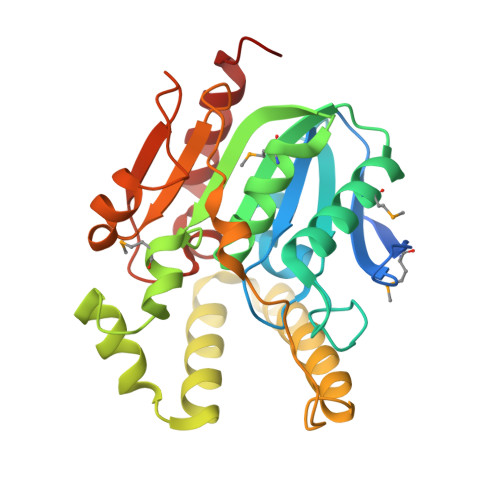
RCSB PDB - 4L9A: Crystal structure of Smu.1393c from cariogenic pathogen Streptococcus mutans

 4L9A

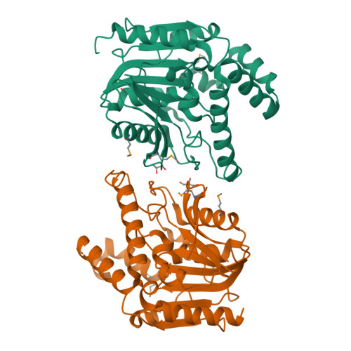
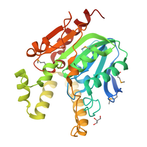
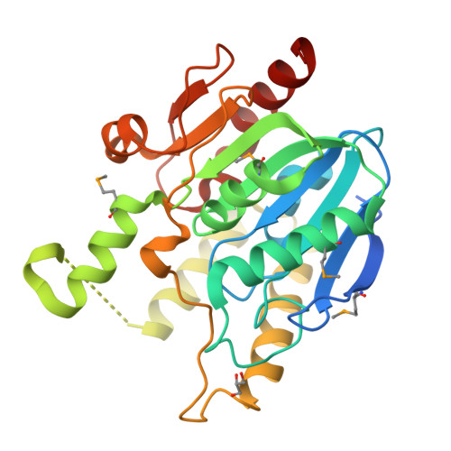
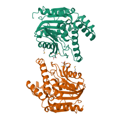
Crystal structure of Smu.1393c from cariogenic pathogen Streptococcus mutans


Experimental Data Snapshot

  • Method: X-RAY DIFFRACTION
  • Resolution: 2.00 Å
  • R-Value Free: 0.205 
  • R-Value Work: 0.165 
  • R-Value Observed: 0.167 

wwPDB Validation   3D Report Full Report


This is version 1.3 of the entry. See complete history


Literature

Structural and functional characterization of a novel alpha / beta hydrolase from cariogenic pathogen Streptococcus mutans.

Wang, Z.Li, L.Su, X.D.

(2014) Proteins 82: 695-700

  • DOI: https://doi.org/10.1002/prot.24418
  • Primary Citation of Related Structures:  
    4L9A

  • PubMed Abstract: 

    The protein Smu.1393c from Streptococcus mutans is annotated as a putative α/β hydrolase, but it has low sequence identity to the structure-known α/β hydrolases. Here we present the crystal structure of Smu.1393c at 2.0 Å resolution. Smu.1393c has a fully open alkaline substrate pocket, whose conformation is unique among other similar hydrolase structures. Three residues, Ser101, His251, and Glu125, were identified as the active center of Smu.1393c. By screening a series of artificial hydrolase substrates, we demonstrated Smu.1393c had low carboxylesterase activity towards short-chain carboxyl esters, which provided a clue for exploring the in vivo function of Smu.1393c.


  • Organizational Affiliation

    State Key Laboratory of Protein and Plant Gene Research, and Biodynamic Optical Imaging Center (BIOPIC), School of Life Sciences, Peking University, Beijing, 100871, China.


Macromolecules
Find similar proteins by:  (by identity cutoff)  |  3D Structure
Entity ID: 1
MoleculeChains Sequence LengthOrganismDetailsImage
Putative uncharacterized protein Smu.1393c
A, B
292Streptococcus mutans UA159Mutation(s): 0 
Gene Names: SMU_1393c
UniProt
Find proteins for Q8DTF4 (Streptococcus mutans serotype c (strain ATCC 700610 / UA159))
Explore Q8DTF4 
Go to UniProtKB:  Q8DTF4
Entity Groups  
Sequence Clusters30% Identity50% Identity70% Identity90% Identity95% Identity100% Identity
UniProt GroupQ8DTF4
Sequence Annotations
Expand
  • Reference Sequence
Small Molecules
Modified Residues  1 Unique
IDChains TypeFormula2D DiagramParent
MSE
Query on MSE
A, B
L-PEPTIDE LINKINGC5 H11 N O2 SeMET
Experimental Data & Validation

Experimental Data

  • Method: X-RAY DIFFRACTION
  • Resolution: 2.00 Å
  • R-Value Free: 0.205 
  • R-Value Work: 0.165 
  • R-Value Observed: 0.167 
  • Space Group: P 1 21 1
Unit Cell:
Length ( Å )Angle ( ˚ )
a = 43.504α = 90
b = 135.958β = 92.51
c = 51.568γ = 90
Software Package:
Software NamePurpose
ADSCdata collection
PHENIXmodel building
PHENIXrefinement
HKL-2000data reduction
HKL-2000data scaling
PHENIXphasing

Structure Validation

View Full Validation Report


Entry History 

Deposition Data

Revision History  (Full details and data files)

  • Version 1.0: 2013-07-17
    Type: Initial release
  • Version 1.1: 2013-11-06
    Changes: Database references
  • Version 1.2: 2022-08-24
    Changes: Database references, Derived calculations
  • Version 1.3: 2024-11-06
    Changes: Data collection, Structure summary