4G5R

Structure of LGN GL4/Galphai3 complex


Experimental Data Snapshot

  • Method: X-RAY DIFFRACTION
  • Resolution: 3.48 Å
  • R-Value Free: 
    0.253 (Depositor), 0.250 (DCC) 
  • R-Value Work: 
    0.209 (Depositor), 0.210 (DCC) 
  • R-Value Observed: 
    0.211 (Depositor) 

wwPDB Validation   3D Report Full Report


Ligand Structure Quality Assessment 

Created with Raphaël 2.3.0Worse 01 BetterLigand structure goodness of fit to experimental dataBest fitted GDPClick on this verticalbar to view details

This is version 1.2 of the entry. See complete history


Literature

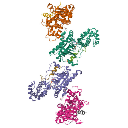
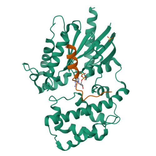
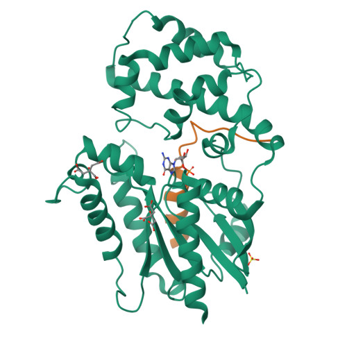
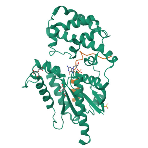
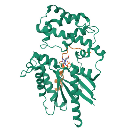
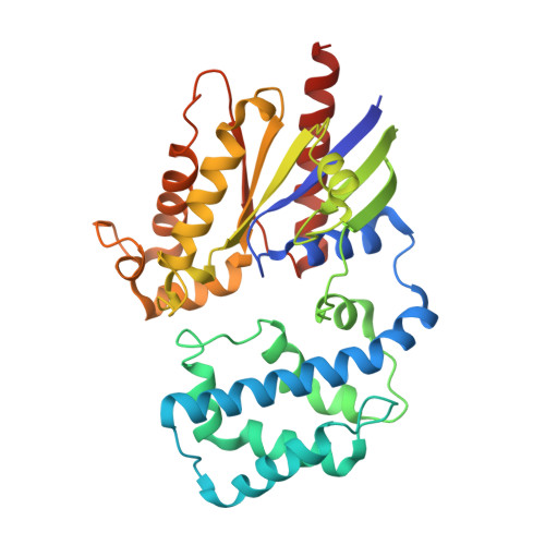
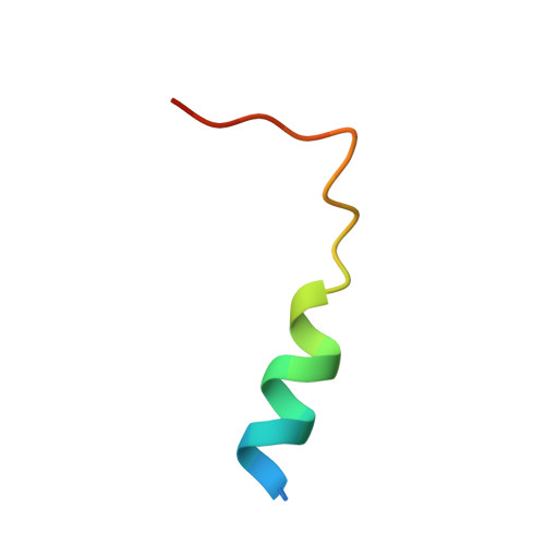
Crystal structures of the scaffolding protein LGN reveal the general mechanism by which GoLoco binding motifs inhibit the release of GDP from G alpha i.

Jia, M.Li, J.Zhu, J.Wen, W.Zhang, M.Wang, W.

(2012) J Biol Chem 287: 36766-36776

  • DOI: https://doi.org/10.1074/jbc.M112.391607
  • Primary Citation of Related Structures:  
    4G5O, 4G5Q, 4G5R, 4G5S

  • PubMed Abstract: 

    GoLoco (GL) motif-containing proteins regulate G protein signaling by binding to Gα subunit and acting as guanine nucleotide dissociation inhibitors. GLs of LGN are also known to bind the GDP form of Gα(i/o) during asymmetric cell division. Here, we show that the C-terminal GL domain of LGN binds four molecules of Gα(i)·GDP. The crystal structures of Gα(i)·GDP in complex with LGN GL3 and GL4, respectively, reveal distinct GL/Gα(i) interaction features when compared with the only high resolution structure known with GL/Gα(i) interaction between RGS14 and Gα(i1.) Only a few residues C-terminal to the conserved GL sequence are required for LGN GLs to bind to Gα(i)·GDP. A highly conserved "double Arg finger" sequence (RΨ(D/E)(D/E)QR) is responsible for LGN GL to bind to GDP bound to Gα(i). Together with the sequence alignment, we suggest that the LGN GL/Gα(i) interaction represents a general binding mode between GL motifs and Gα(i). We also show that LGN GLs are potent guanine nucleotide dissociation inhibitors.


  • Organizational Affiliation

    Shanghai Key Laboratory of Molecular Catalysis and Innovative Materials, Department of Chemistry and Institute of Biomedical Sciences, Fudan University, Shanghai 200433, China.


Macromolecules
Find similar proteins by:  (by identity cutoff)  |  3D Structure
Entity ID: 1
MoleculeChains Sequence LengthOrganismDetailsImage
Guanine nucleotide-binding protein G(k) subunit alpha
A, B, C, D
330Homo sapiensMutation(s): 0 
Gene Names: GNAI3
UniProt & NIH Common Fund Data Resources
Find proteins for P08754 (Homo sapiens)
Explore P08754 
Go to UniProtKB:  P08754
PHAROS:  P08754
GTEx:  ENSG00000065135 
Entity Groups  
Sequence Clusters30% Identity50% Identity70% Identity90% Identity95% Identity100% Identity
UniProt GroupP08754
Sequence Annotations
Expand
  • Reference Sequence

Find similar proteins by:  Sequence   |   3D Structure  

Entity ID: 2
MoleculeChains Sequence LengthOrganismDetailsImage
G-protein-signaling modulator 2E,
F,
G,
H [auth Z]
25Mus musculusMutation(s): 0 
UniProt & NIH Common Fund Data Resources
Find proteins for Q8VDU0 (Mus musculus)
Explore Q8VDU0 
Go to UniProtKB:  Q8VDU0
IMPC:  MGI:1923373
Entity Groups  
Sequence Clusters30% Identity50% Identity70% Identity90% Identity95% Identity100% Identity
UniProt GroupQ8VDU0
Sequence Annotations
Expand
  • Reference Sequence
Small Molecules
Ligands 3 Unique
IDChains Name / Formula / InChI Key2D Diagram3D Interactions
GDP
Query on GDP

Download Ideal Coordinates CCD File 
I [auth A],
K [auth B],
O [auth C],
S [auth D]
GUANOSINE-5'-DIPHOSPHATE
C10 H15 N5 O11 P2
QGWNDRXFNXRZMB-UUOKFMHZSA-N
CIT
Query on CIT

Download Ideal Coordinates CCD File 
L [auth B],
M [auth B],
P [auth C],
Q [auth C]
CITRIC ACID
C6 H8 O7
KRKNYBCHXYNGOX-UHFFFAOYSA-N
SO4
Query on SO4

Download Ideal Coordinates CCD File 
J [auth A],
N [auth B],
R [auth C]
SULFATE ION
O4 S
QAOWNCQODCNURD-UHFFFAOYSA-L
Experimental Data & Validation

Experimental Data

  • Method: X-RAY DIFFRACTION
  • Resolution: 3.48 Å
  • R-Value Free:  0.253 (Depositor), 0.250 (DCC) 
  • R-Value Work:  0.209 (Depositor), 0.210 (DCC) 
  • R-Value Observed: 0.211 (Depositor) 
Space Group: P 61 2 2
Unit Cell:
Length ( Å )Angle ( ˚ )
a = 209.55α = 90
b = 209.55β = 90
c = 237.211γ = 120
Software Package:
Software NamePurpose
REFMACrefinement
PDB_EXTRACTdata extraction
HKL-2000data collection
HKL-2000data reduction
HKL-2000data scaling
MOLREPphasing
PHENIXrefinement

Structure Validation

View Full Validation Report


Ligand Structure Quality Assessment 

Created with Raphaël 2.3.0Worse 01 BetterLigand structure goodness of fit to experimental dataBest fitted GDPClick on this verticalbar to view details

Entry History 

Deposition Data

Revision History  (Full details and data files)

  • Version 1.0: 2012-09-05
    Type: Initial release
  • Version 1.1: 2024-03-20
    Changes: Data collection, Database references, Derived calculations
  • Version 1.2: 2025-03-05
    Changes: Database references, Structure summary