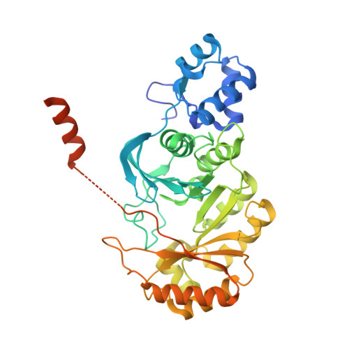
3RF9

X-ray structure of RlmN from Escherichia coli


Experimental Data Snapshot

  • Method: X-RAY DIFFRACTION
  • Resolution: 2.20 Å
  • R-Value Free: 0.267 
  • R-Value Work: 0.213 
  • R-Value Observed: 0.216 

wwPDB Validation   3D Report Full Report


This is version 1.3 of the entry. See complete history


Literature

Structural basis for methyl transfer by a radical SAM enzyme.

Boal, A.K.Grove, T.L.McLaughlin, M.I.Yennawar, N.H.Booker, S.J.Rosenzweig, A.C.

(2011) Science 332: 1089-1092

  • DOI: https://doi.org/10.1126/science.1205358
  • Primary Citation of Related Structures:  
    3RF9, 3RFA

  • PubMed Abstract: 

    The radical S-adenosyl-L-methionine (SAM) enzymes RlmN and Cfr methylate 23S ribosomal RNA, modifying the C2 or C8 position of adenosine 2503. The methyl groups are installed by a two-step sequence involving initial methylation of a conserved Cys residue (RlmN Cys(355)) by SAM. Methyl transfer to the substrate requires reductive cleavage of a second equivalent of SAM. Crystal structures of RlmN and RlmN with SAM show that a single molecule of SAM coordinates the [4Fe-4S] cluster. Residue Cys(355) is S-methylated and located proximal to the SAM methyl group, suggesting the SAM that is involved in the initial methyl transfer binds at the same site. Thus, RlmN accomplishes its complex reaction with structural economy, harnessing the two most important reactivities of SAM within a single site.


  • Organizational Affiliation

    Department of Molecular Biosciences, Northwestern University, Evanston, IL 60208, USA.


Macromolecules
Find similar proteins by:  (by identity cutoff)  |  3D Structure
Entity ID: 1
MoleculeChains Sequence LengthOrganismDetailsImage
Ribosomal RNA large subunit methyltransferase N
A, B
404Escherichia coli K-12Mutation(s): 0 
Gene Names: rlmNyfgBb2517JW2501
EC: 2.1.1.192
UniProt
Find proteins for P36979 (Escherichia coli (strain K12))
Go to UniProtKB:  P36979
Sequence Annotations
Expand
  • Reference Sequence
Experimental Data & Validation

Experimental Data

  • Method: X-RAY DIFFRACTION
  • Resolution: 2.20 Å
  • R-Value Free: 0.267 
  • R-Value Work: 0.213 
  • R-Value Observed: 0.216 
  • Space Group: C 2 2 21
Unit Cell:
Length ( Å )Angle ( ˚ )
a = 72.152α = 90
b = 80.406β = 90
c = 312.242γ = 90
Software Package:
Software NamePurpose
DENZOdata reduction
SCALEPACKdata scaling
REFMACrefinement
PDB_EXTRACTdata extraction
HKL-2000data collection
SHARPphasing

Structure Validation

View Full Validation Report


Entry History 

Deposition Data

Revision History  (Full details and data files)

  • Version 1.0: 2011-05-11
    Type: Initial release
  • Version 1.1: 2011-07-13
    Changes: Version format compliance
  • Version 1.2: 2017-11-08
    Changes: Refinement description
  • Version 1.3: 2024-02-21
    Changes: Data collection, Database references, Derived calculations